. 2010 Sep-Oct;57(5):405-14.
doi: 10.1111/j.1550-7408.2010.00496.x. Epub 2010 Aug 5.

Evidence for rosettes as an unrecognized stage in the life cycle of Leishmania parasites

Affiliations

Evidence for rosettes as an unrecognized stage in the life cycle of Leishmania parasites

David M Iovannisci et al. J Eukaryot Microbiol. 2010 Sep-Oct.

Abstract

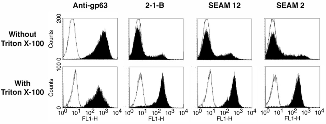
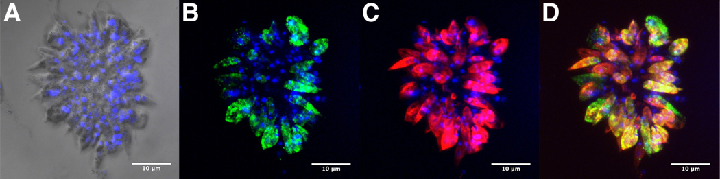
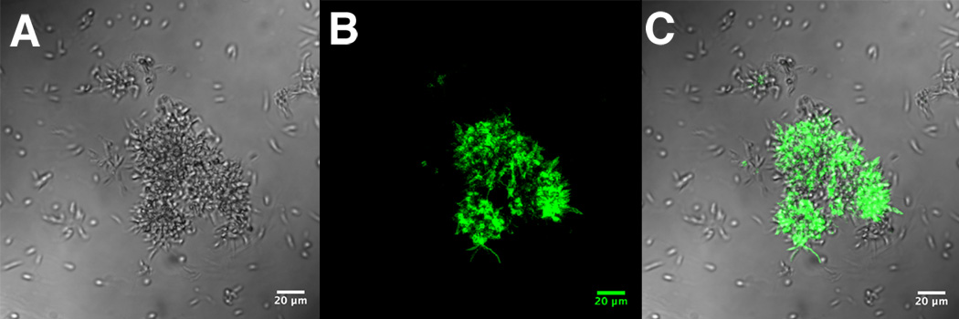
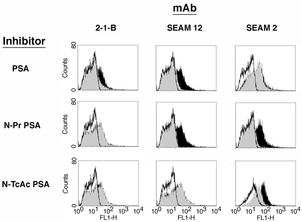
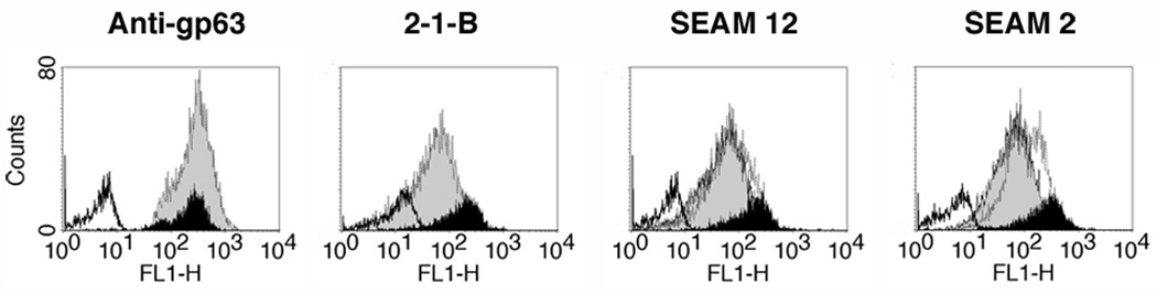
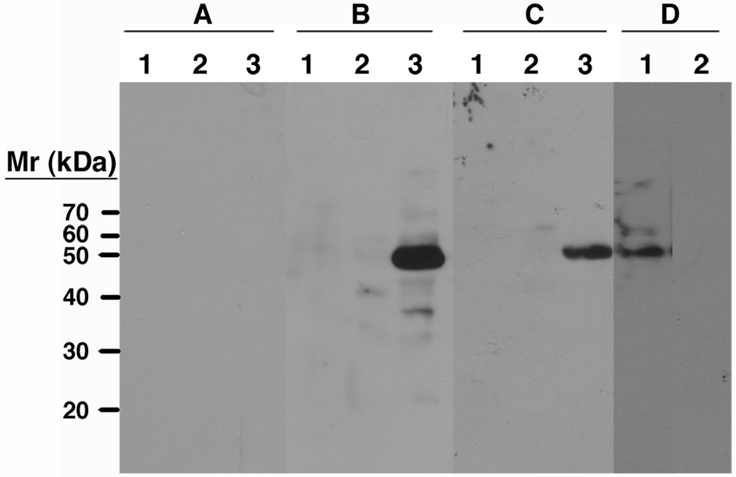
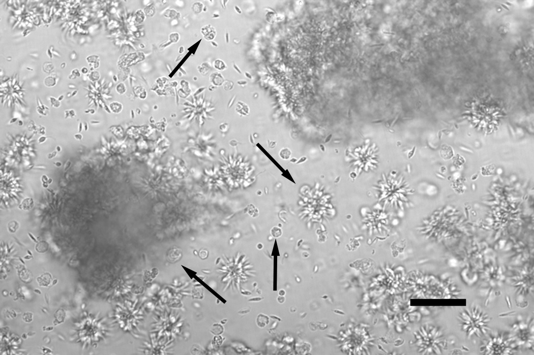
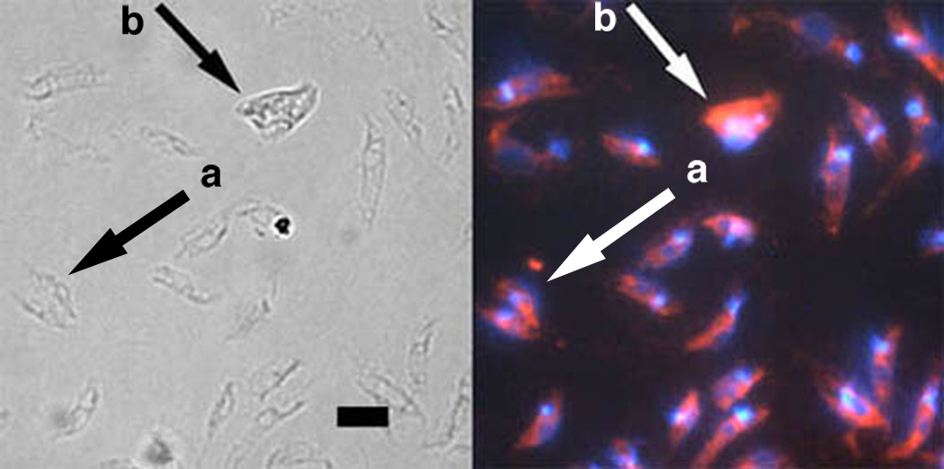
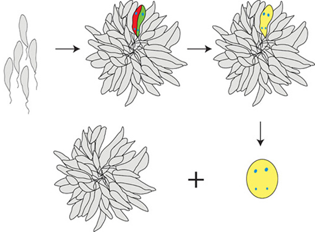
Leishmania parasites, which afflict 12 million people in 88 countries, exist as promastigotes transmitted by insect vectors and as amastigotes residing in mammalian macrophages. Promastigote cells arranged in rosettes have also been described but universally disregarded as a distinct stage in the life cycle. We present evidence that only rosettes of Leishmania major promastigotes express cell surface poly-alpha2,8 N-acetyl neuraminic acid (PSA) and PSA containing de-N-acetyl neuraminic acid (NeuPSA). Expression of rosette-specific PSA antigens was mosaic, with individual promastigotes expressing PSA, NeuPSA or both. A 50 kDa protein was detected by Western blot analysis of a detergent-insoluble cell fraction with both PSA and NeuPSA-reactive antibodies. Frequencies of rosette formation as well as cell surface PSA/NeuPSA expression were temperature dependent. Rosettes also engaged in an unusual swarming behavior, congregating into extended clusters. Distinct structures resembling cellular fusion bodies were formed in and released from rosettes. The results indicate that rosettes are an unrecognized stage in the life cycle of Leishmania. We hypothesize that rosettes initiate mating in Leishmania during which PSA/NeuPSA expression plays an important role. Recognizing rosettes as a distinct form of the Leishmania life cycle opens new possibilities for treatment or prevention of disease and, possibly, in vitro genetic recombination without passage of cells through insect vectors.


Figures

Fig. 1
Fig. 1
Detection by monoclonal antibodies (mAb) of Poly α 2,8 N-acetyl neuraminic acid (PSA) and PSA containing de-N-acetyl neuraminic acid (NeuPSA) in Leishmania major. Promastigote cells were labeled with each mAb indicated and examined by flow cytometry. Upper panel, binding without Triton X-100 exposure to detect only surface-associated antigens; lower panel, mAb binding after a 10 min Triton X-100 exposure to detect both cytoplasmic and surface-associated antigens. Black fill, binding of indicated mAb; white fill, irrelevant isotype matched control mAbs. Results indicate that nearly all leishmanial promastigotes possess PSA/NeuPSA internally, but only a small fraction of the population expresses these antigens on their surface when grown at 24--26 °C. In contrast, nearly all cells express cell surface gp63 under the same culture conditions.
Fig. 2
Fig. 2
Confocal microscopic examination of Leishmania major rosettes labeled with Poly α 2,8 N-acetyl neuraminic acid (PSA) and PSA containing de-N-acetyl neuraminic acid (NeuPSA). A, light micrograph with DAPI-fluorescent labeling overlay; B, anti-PSA mAb 2-1-B/DAPI labeling; C, anti-NeuPSA mAb SEAM 2/DAPI labeling; D, composite of mAb 2-1-B/mAb SEAM 2/DAPI labeling. Individual promastigote cells within the rosette expressing PSA, NeuPSA or both can be observed. Scale bars, 10 µm.
Fig. 3
Fig. 3
Confocal microscopic examination of Leishmania major culture containing a mixture of rosettes and individual promastigotes labeled with the anti-Poly α 2,8 N-acetyl neuraminic acid (PSA) mAb 2-1-B. A, light micrograph of a mixture of individual promastigotes and promastigotes within rosettes; B, PSA labeling of promastigotes shown in A with 2-1-B; C, composite of A and B. Only promastigote cells within rosettes express PSA. Scale bars, 20 µm.
Fig. 4
Fig. 4
Specific inhibition of anti-Poly α 2,8 N-acetyl neuraminic acid (anti-PSA) and anti-PSA containing de-N-acetyl neuraminic acid (anti-NeuPSA) mAb binding to leishmanial rosettes. Monoclonal antibodies (mAbs) were preincubated with each inhibitor for 20 min and subsequently added to rosette preparations. Poly α 2,8 N-acetyl neuraminic acid (PSA) is reactive with mAbs 2-1-B and SEAM 12 but not mAb SEAM 2; N-propionyl PSA (N-Pr PSA) is reactive with mAbs SEAM 12 and SEAM 2 but not mAb 2-1-B; N-trichloroacetyl PSA (N-TcAc PSA) is reactive only with mAb SEAM 2. Labeled cells were examined by flow cytometry. Black fill, indicates mAb binding; gray fill, indicates mAb signal in the presence of specific inhibitor; white fill, irrelevant isotype matched control mAb. Specific inhibition of each anti-PSA and anti-NeuPSA mAb is observed.
Fig. 5
Fig. 5
Temperature dependency of cell surface expression of Poly α 2,8 N-acetyl neuraminic acid (PSA) and PSA containing de-N-acetyl neuraminic acid (NeuPSA) by leishmanial rosettes. Cells were cultivated at 16 °C (dotted line, white fill), 20 °C (black fill), and 24 °C (dotted line, gray fill) for at least 4 weeks prior to labeling. Irrelevant isotype matched control mAb (solid line, white fill, signal intensity ≤10). Maximal cell surface expression of PSA/NeuPSA occurs when promastigotes were cultured at 20 °C.
Fig. 6
Fig. 6
Western blot of Triton X-114 fractionated leishmanial rosette preparations. A, irrelevant mAb control; B, mAb SEAM 12; C, mAb SEAM 2. Lane 1, Triton X-114 fraction; lane 2, buffer fraction; lane 3, detergent insoluble cell pellet fraction; D, detergent insoluble cell pellet fraction with SEAM 12 in the absence (lane 1) or presence (lane 2) of 100 µg/ml N-Pr PSA as a specific inhibitor of SEAM 12 binding. A single band of approximately 50kDa from the detergent insoluble cell pellet is identified by anti-PSA/NeuPSA and its detection specifically inhibited by N-Pr PSA.
Fig. 7
Fig. 7
Observation of rosette swarming in Leishmania. A, 0 h; B, 2 h; C, 4 h; D, 8 h; E, 30 h. At 0 h, a confluent cell suspension is observable. At 2 h, a spotty appearance is detectable corresponding to regions of higher and lower cell density. These elongate at 4 h and begin to converge at 8 h. Cells eventually congregate into a star-like appearance in the center of the Petri dish.
Fig. 8
Fig. 8
Generation of fusion bodies from leishmanial rosettes. Micrograph taken of a live, unmanipulated culture. Individual as well as large aggregates of rosettes can be seen. Center downward arrow indicates 2 spherical fusion bodies forming on a single rosette. Additional arrows indicate numerous individual fusion bodies that have been released from rosettes. Note the size and morphology compared to nearby promastigote cells. These morphologies are distinct from any previously described leishmanial cellular structures, such as amastigotes, promastigotes or rosettes. Scale bar: 50 µm.
Fig. 9
Fig. 9
Labeling of fusion bodies by Leishmania specific anti–gp63 monoclonal antibody (mAb). Left panel, light microscopy; right panel, fluorescence microscopy. Numerous individual promastigotes are visible and contain a single nucleus and single kinetoplast stained with DAPI. A dividing cell (arrow a) and fusion body (arrow b) are shown along with individual promatigotes. All cells including the fusion body were detected by the Leishmania specific anti–gp63 mAb, demonstrating that fusion bodies are not artifacts or contaminants. Fusion bodies contain multiple DAPI staining foci as expected by the fusion of two or more leishmanial promastigotes. The ratio of the integrated pixel density of DAPI DNA staining in the dividing cell and fusion body compared to five individual promastigotes in the same micrograph were 1.88 and 2.70, respectively. Scale bar, 10 µm.
Fig. 10
Fig. 10
Proposed model for the role of rosette formation, and of Poly α 2,8 N-acetyl neuraminic acid (PSA) and PSA containing de-N-acetyl neuraminic acid (NeuPSA) expression in the leishmanial life cycle. Under the appropriate growth conditions, promastigotes of Leishmania form rosettes in significant numbers and begin to express cell surface PSA and NeuPSA. For simplicity only two cells within a single rosette are shown expressing PSA or NeuPSA (labeled red and green, respectively), each containing a single nucleus and kinetoplast labeled in blue. Cell surface PSA/NeuPSA expression promotes the fusion of two or more neighboring promastigotes (labeled yellow), allowing the potential co-mingling of their kinetoplast and nuclear genomes. Fusion bodies, subsequently exhibit a distinct morphology, and are released from the rosettes.

Similar articles

Cited by

References

    1. Abramoff MD, Magelhaes PJ, Ram SJ. Image processing with ImageJ. Biophotonics Internl. 2004;11:36–42.
    1. Adair WS, Monk BC, Cohen R, Hwang C, Goodenough UW. Sexual agglutinins from the Chlamydomonas flagellar membrane. Partial purification and characterization. J. Biol. Chem. 1982;257:4593–4602. - PubMed
    1. Akopyants NS, Kimblin N, Secundino N, Patrick R, Peters N, Lawyer P, Dobson DE, Beverley SM, Sacks DL. Demonstration of genetic exchange during cyclical development of Leishmania in the sand fly vector. Science. 2009;324:265–268. - PMC - PubMed
    1. Angata T, Varki A. Chemical diversity in the sialic acids and related alpha-keto acids: An evolutionary perspective. Chem. Rev. 2002;102:439–469. - PubMed
    1. Bastien P, Blaineau C, Pages M. Leishmania: sex, lies and karyotype. Parasitol. Today. 1992;8:174–177. - PubMed

Publication types

MeSH terms

Substances