. 2010 Nov 12;402(2):180-5.
doi: 10.1016/j.bbrc.2010.08.022. Epub 2010 Aug 11.

T3 affects expression of collagen I and collagen cross-linking in bone cell cultures

Affiliations

T3 affects expression of collagen I and collagen cross-linking in bone cell cultures

F Varga et al. Biochem Biophys Res Commun. .

Abstract

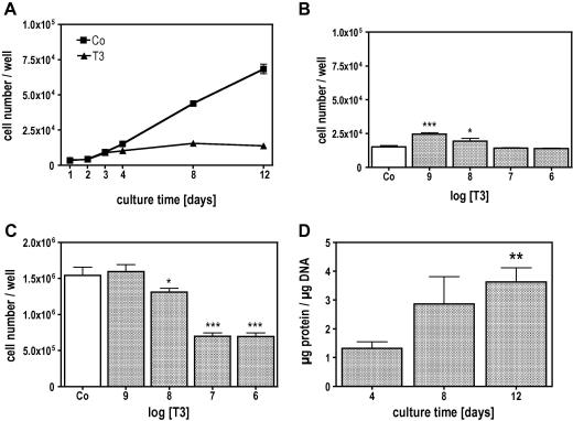
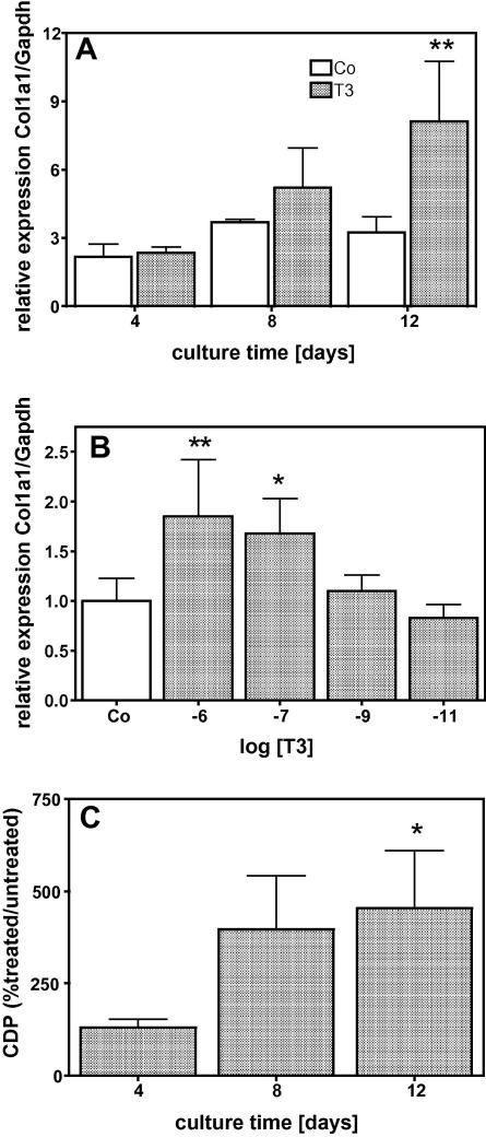
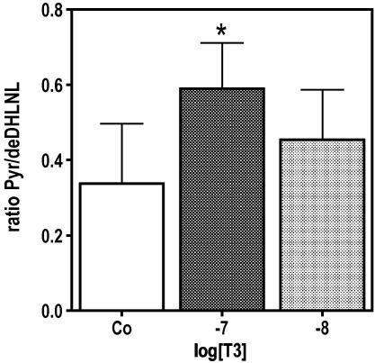
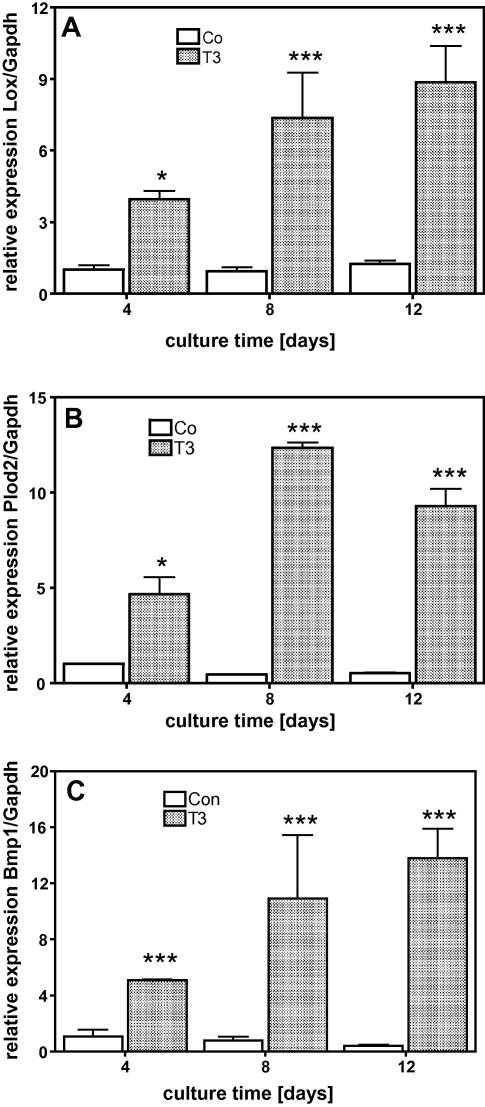
Thyroid hormones (T3,T4) have a broad range of effects on bone, however, its role in determining the quality of bone matrix is poorly understood. In-vitro, the immortalized mouse osteoblast-like cell line MC3T3-E1 forms a tissue like structure, consisting of several cell layers, whose formation is affected by T3 significantly. In this culture system, we investigated the effects of T3 on cell multiplication, collagen synthesis, expression of genes related to the collagen cross-linking process and on the formation of cross-links. T3 compared to controls modulated cell multiplication, up-regulated collagen synthesis time and dose dependently, and stimulated protein synthesis. T3 increased mRNA expressions of procollagen-lysine-1,2-oxoglutarate 5-dioxygenase 2 (Plod2) and of lysyloxidase (Lox), both genes involved in post-translational modification of collagen. Moreover, it stimulated mRNA expression of bone morphogenetic protein 1 (Bmp1), the processing enzyme of the lysyloxidase-precursor and of procollagen. An increase in the collagen cross-link-ratio Pyr/deDHLNL indicates, that T3 modulated cross-link maturation in the MC3T3-E1 culture system. These results demonstrate that T3 directly regulates collagen synthesis and collagen cross-linking by up-regulating gene expression of the specific cross-link related enzymes, and underlines the importance of a well-balanced concentration of thyroid hormones for maintenance of bone quality.


Figures

Fig. 1
Fig. 1
T3 regulated cell multiplication and protein synthesis of MC3T3-E1 osteoblastic cells. Cells were cultured in absence (Co) or presence of T3 (10−7 M) for the indicated time. During culture T3 attenuated cell multiplication beginning with day 4 (A) significantly (P ⩽ 0.001). The effect of T3 was dose dependent. T3 significantly increased cell multiplication at 10−9 and 10−8 M on day 4 (B) while on day 12 (C) T3 attenuated cell multiplication at 10−8, 10−7 and 10−6 M, significantly. On day 12, T3 increased protein synthesis significantly when normalized to the DNA-amount. The bars indicate mean ± SD. ∗P ⩽ 0.05; ∗∗P ⩽ 0.01; ∗∗∗P ⩽ 0.001; Co vs. treatments (n = 4).
Fig. 2
Fig. 2
T3 stimulated collagen α1 (I) expression in MC3T3-E1 osteoblastic cells time and dose dependently. Cells were cultured in absence (Co) or presence of 10−7 M T3 for 4, 8 and 12 days (A) and on day 12 with increasing concentrations T3 (B). T3 time and dose dependently increased Col1a1 mRNA expression reaching significance on day 12 (A) at 10−7 and 10−6 M (B). (C) Up-regulation of Col1a1 mRNA was translated into protein as shown by collagenase digestible protein (CDP) that was estimated as a surrogate for collagen protein synthesis. The bars indicate mean ± SD. ∗P ⩽ 0.05; ∗∗P ⩽ 0.01; Co vs. treatments (n = 4).
Fig. 3
Fig. 3
T3 modify cross-link of the ECM produced by MC3T3-E1 cells. MC3T3-E1 cells were cultured in absence (Co) or presence of 10−8 and 10−7 M T3 for 12 days. Thereafter, cell layers were fixed with 70% Ethanol and specific cross-links were analysed by FTIR. T3 increased cross-link ratio (Pyr/deDHLNL) as a function of concentration, which showed significant difference at 10−7 M T3. Provided P-values indicate significantly different values vs. control cultures. The bars indicate mean ± SD. ∗P ⩽ 0.05 (n = 3).
Fig. 4
Fig. 4
T3 stimulated mRNA expression of genes related to collagen cross-linking process. Cells were cultured in absence (Co) or presence of 10−7 M T3 for 4, 8 and 12 days. Compared to untreated controls T3 increased expression of Lox (A), Plod2 (B) and Bmp1 (C) significantly. Total RNA was isolated and subjected to QRT-PCR, which was performed as triplicate. The bars indicate mean ± SD of a typical experiment. ∗P ⩽ 0.05; ∗∗∗P ⩽ 0.001 (n = 3).

Similar articles

Cited by

References

    1. Murphy E., Williams G.R. The thyroid and the skeleton. Clin. Endocrinol. (Oxf.) 2004;61:285–298. - PubMed
    1. Allain T.J., McGregor A.M. Thyroid hormones and bone. J. Endocrinol. 1993;139:9–18. - PubMed
    1. Gothe S., Wang Z., Ng L., Kindblom J.M., Barros A.C., Ohlsson C., Vennstrom B., Forrest D. Mice devoid of all known thyroid hormone receptors are viable but exhibit disorders of the pituitary–thyroid axis, growth, and bone maturation. Genes Dev. 1999;13:1329–1341. - PMC - PubMed
    1. Nilsson O., Marino R., De Luca F., Phillip M., Baron J. Endocrine regulation of the growth plate. Horm. Res. 2005;64:157–165. - PubMed
    1. Harvey C.B., O’Shea P.J., Scott A.J., Robson H., Siebler T., Shalet S.M., Samarut J., Chassande O., Williams G.R. Molecular mechanisms of thyroid hormone effects on bone growth and function. Mol. Genet. Metab. 2002;75:17–30. - PubMed

Publication types