. 2010 Aug 9;18(2):147-59.
doi: 10.1016/j.ccr.2010.06.015.

Phosphorylation by casein kinase I promotes the turnover of the Mdm2 oncoprotein via the SCF(beta-TRCP) ubiquitin ligase

Affiliations

Phosphorylation by casein kinase I promotes the turnover of the Mdm2 oncoprotein via the SCF(beta-TRCP) ubiquitin ligase

Hiroyuki Inuzuka et al. Cancer Cell. .

Abstract

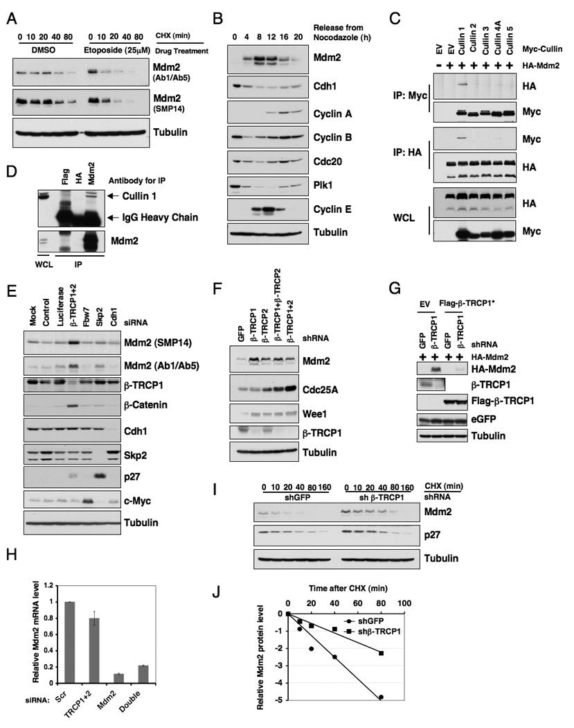
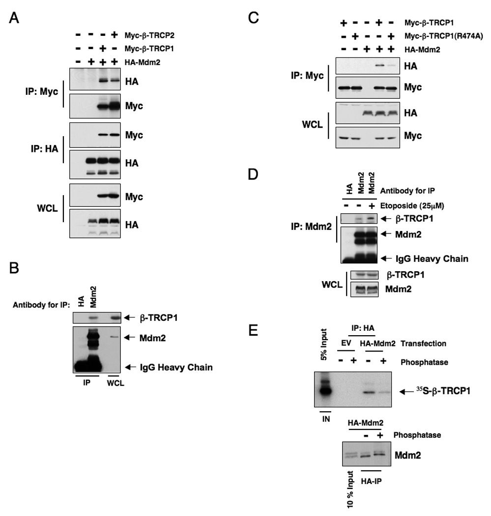
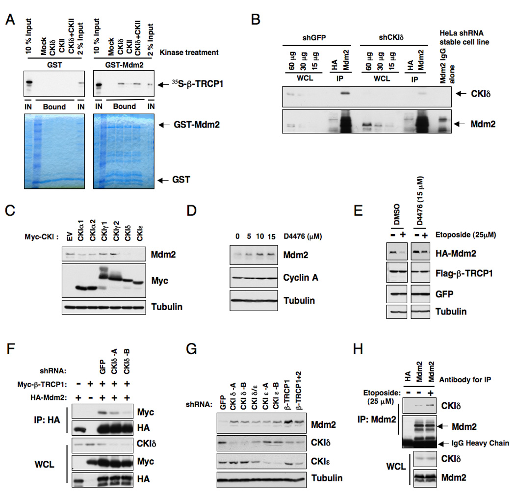
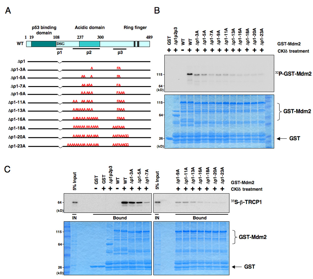
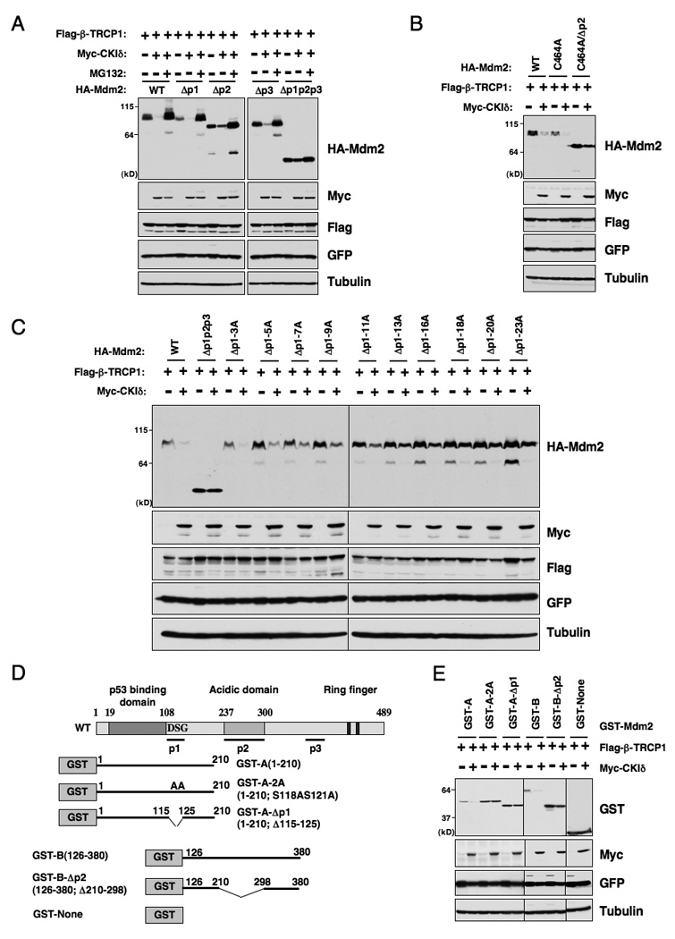
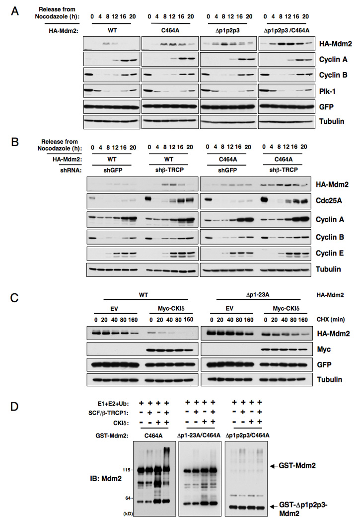
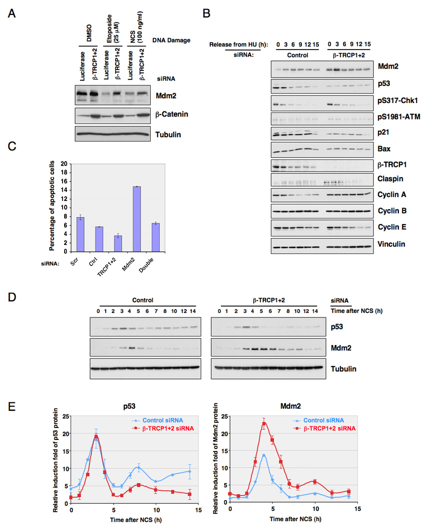
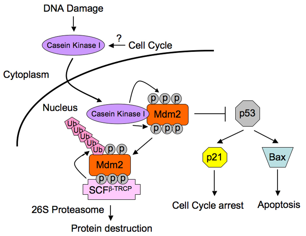
Mdm2 is the major negative regulator of the p53 pathway. Here, we report that Mdm2 is rapidly degraded after DNA damage and that phosphorylation of Mdm2 by casein kinase I (CKI) at multiple sites triggers its interaction with, and subsequent ubiquitination and destruction, by SCF(beta-TRCP). Inactivation of either beta-TRCP or CKI results in accumulation of Mdm2 and decreased p53 activity, and resistance to apoptosis induced by DNA damaging agents. Moreover, SCF(beta-TRCP)-dependent Mdm2 turnover also contributes to the control of repeated p53 pulses in response to persistent DNA damage. Our results provide insight into the signaling pathways controlling Mdm2 destruction and further suggest that compromised regulation of Mdm2 results in attenuated p53 activity, thereby facilitating tumor progression.


Figures

Figure 1
Figure 1. Mdm2 stability is controlled by β-TRCP
A. U2OS cells were treated with 25 µM etoposide for 1.5 hours before 20 µg/ml cycloheximide was added. At the indicated time points, whole cell lysates were prepared and immunoblots were probed with the indicated antibodies. B. Immunoblot analysis of HeLa cells synchronized by growth in nocodazole, and then released for the indicated periods of time. C. Immunoblot analysis of whole cell lysates (WCL) and immunoprecipitates (IP) derived from 293T cells transfected with HA-Mdm2 and various Myc-tagged Cullin constructs. Twenty hours post-transfection, cells were treated with 10 µM MG132 overnight before harvesting. D. Immunoblot analysis of HeLa cell whole cell lysates (WCL) and anti-Mdm2 immunoprecipitates (IP). Flag- and HA-agarose beads were used as negative controls for the immunoprecipitation procedure. Cells were treated with 10 µM MG132 overnight before harvesting. E. Immunoblot analysis of HeLa cells transfected with the indicated siRNA oligonucleotides. The control lane is scrambled E2F-1 siRNA; Luciferase, siRNA against firefly luciferase; β-TRCP1+2, siRNA oligo that can deplete both β-TRCP1 and β-TRCP2 isoforms. siRNA, short interfering RNA. F. Immunoblot analysis of HeLa cells transfected with the indicated shRNA constructs. G. Immunoblot analysis of 293T cells transfected with the indicated shRNA constructs together with HA-Mdm2 and jellyfish GFP (eGFP, as transfection controls). Where indicated, Flag-β-TRCP1*, which is engineered to resist the shβ-TRCP1 effect, was included in the transfection. H. Real-time RT-PCR analysis to examine the relative Mdm2 mRNA expression levels in U2OS cells transfected with the indicated siRNA oligonucleotides. Three independent sets of experiments were performed to generate the error bar. The error bars represent +/− SD. I. HeLa cells were transfected with the indicated shRNA constructs. Twenty hours post-transfection, cells were split into 60 mm dishes, and after another 20 hours, treated with 20 µg/ml cycloheximide. At the indicated time points, whole cell lysates were prepared and immunoblots were probed with the indicated antibodies. J. Quantification of the band intensities in I. Mdm2 band intensity was normalized to tubulin, then normalized to the t=0 controls. See also Figure S1.
Figure 2
Figure 2. Mdm2 interacts with β-TRCP in a phosphorylation dependent manner
A. Immunoblot analysis of whole cell lysates (WCL) and immunoprecipitates (IP) derived from 293T cells transfected with HA-Mdm2 and Myc-tagged β-TRCP constructs. Twenty hours post-transfection, cells were treated with 10 µM MG132 overnight before harvesting. B. Immunoblot analysis of HeLa cell whole cell lysates (WCL) and anti-Mdm2 immunoprecipitates (IP). HA-agarose beads were used as a negative control for the IP. Cells were treated with 10 µM MG132 overnight before harvesting. C. Immunoblot analysis of whole cell lysates (WCL) and immunoprecipitates (IP) derived from 293T cells transfected with HA-Mdm2 and the indicated Myc-tagged β-TRCP constructs. Twenty hours post-transfection, cells were treated with 10 µM MG132 overnight before harvesting. D. Immunoblot analysis of HeLa cell whole cell lysates (WCL) and anti-Mdm2 immunoprecipitates (IP). HA-agarose beads were used as a negative control for the IP. Cells were treated with 10 µM MG132 overnight before harvesting. Where indicated, cells were treated with 25 µM etoposide (or DMSO as a negative control) for 30 minutes before harvesting. E. Autoradiograms showing recovery of 35S-labeled β-TRCP1 protein bound to Mdm2 protein immunoprecipitated from HeLa cells transfected with the indicated HA-Mdm2 constructs. Where indicated, the whole cell lysates were treated with λ-phosphatase prior to immunoprecipitation. IN, input (10% or 5% as indicated). See also Figure S2
Figure 3
Figure 3. Casein Kinase I is involved in regulating Mdm2 stability
A. Autoradiograms showing recovery of 35S-labeled β-TRCP1 protein bound to GST-Mdm2 fusion proteins (GST protein as a negative control) incubated with the indicated kinase prior to the pull-down assays. IN, input (10% or 2% as indicated). 35S labeling was carried out by in vitro translation reaction with retic lysate. B. Immunoblot analysis of whole cell lysates (WCL) and anti-Mdm2 immunoprecipitates (IP) derived from the indicated HeLa stable cell lines generated by infection with shGFP or shCKIδ lentiviral construct and subsequent selection with 1 µM puromycin to eliminate the non-infected cells. HA-agarose beads were used as a negative control for the immunoprecipitation procedure. Cells were treated with 10 µM MG132 overnight before harvesting. C. Immunoblot analysis of whole cell lysates (WCL) derived from 293T cells transfected with various Myc-tagged CKI constructs to detect the changes in endogenous Mdm2 expression. D. Immunoblot analysis of HeLa cells treated with the CKI inhibitor D4476 at the indicated concentrations for 12 hours. E. HeLa cells were transiently transfected with the HA-Mdm2 plasmid together with Flag-β-TRCP1. Twenty-four hours post-transfection, cells were treated with the indicated CKI inhibitor for 4 hours and then incubated with 25 µM etoposide for an additional 1.5 hours. The whole cell lysates were recovered and immunoblots were performed with the indicated antibodies. F. Immunoblot analysis of whole cell lysates (WCL) and immunoprecipitates (IP) derived from 293T cells transfected with HA-Mdm2 and Myc-tagged β-TRCP constructs. Where indicated, the shGFP or shCKIδ construct was included in the transfection. Twenty hours post-transfection, cells were treated with 10 µM MG132 overnight before harvesting. G. Immunoblot analysis of HeLa cells transfected with the indicated shRNA constructs. H. Immunoblot analysis of HeLa cell whole cell lysates (WCL) and anti-Mdm2 immunoprecipitates (IP). HA-agarose beads were used as a negative control for the IP. Cells were treated with 10 µM MG132 overnight before harvesting. Where indicated, cells were treated with 25 µM etoposide (or DMSO as control) for 30 minutes before harvesting. See also Figure S3.
Figure 4
Figure 4. Casein Kinase Iδ phosphorylates Mdm2 at multiple sites to trigger Mdm2 interaction with β-TRCP1
A. Illustration of the various Mdm2 mutants generated for this study. B. Sequential inactivation of the potentially critical Ser/Thr sites for individual suboptimal degrons including the identified major CKIδ phosphorylation sites leads to progressive reduction of in vitro Mdm2 phosphorylation signals. Purified CKIδ protein (from New England Biolabs) was incubated with 5 µg of the indicated GST-Mdm2 proteins in the presence of γ-32P-ATP. The kinase reaction products were resolved by SDS-PAGE and phosphorylation was detected by autoradiography. C. Consequently, the recovered in vitro β-TRCP1/Mdm2 interaction is reduced in a step-wise manner after sequential inactivation of the identified Ser/Thr phosphorylation sites in Mdm2, indicating that phosphorylation of Mdm2 at multiple sites by CKIδ triggers its interaction with β-TRCP1 in vitro. Autoradiograms showing recovery of 35S-labeled β-TRCP1 protein bound to the indicated GST-Mdm2 fusion proteins (GST protein as a negative control) incubated with CKIδ prior to the pull-down assays. IN, input (5% as indicated). See also Figure S4.
Figure 5
Figure 5. Casein Kinase Iδ phosphorylates Mdm2 at multiple sites to trigger Mdm2 destruction by β-TRCP1
A. Immunoblot analysis of HeLa cells transfected with the indicated HA-Mdm2 and Flag-β-TRCP1 plasmids in the presence or absence of Myc-CKIδ. A plasmid encoding GFP was used as a negative control for transfection efficiency. Where indicated, the proteasome inhibitor MG132 (10 µM) was added. B. Immunoblot analysis of HeLa cells transfected with various HA-Mdm2C464A constructs (with wild-type Mdm2 as a positive control) and Flag-β-TRCP1 plasmids in the presence or absence of Myc-CKIδ. A plasmid encoding GFP was used as a negative control for transfection efficiency. C. Immunoblot analysis of HeLa cells transfected with the indicated HA-Mdm2 and Flag-β-TRCP1 plasmids in the presence or absence of Myc-CKIδ. A plasmid encoding GFP was used as a negative control for transfection efficiency. D. Illustration of the various GST-Mdm2 constructs used in E and Figure S5. E. Immunoblot analysis of HeLa cells transfected with the indicated GST-Mdm2 and Flag-β-TRCP1 plasmids in the presence or absence of Myc-CKIδ. A plasmid encoding GFP was used as a negative control for transfection efficiency. See also Figure S5.
Figure 6
Figure 6. β-TRCP controls Mdm2 protein abundance during the cell cycle progression, and promotes the ubiquitination of the Mdm2 protein
A. Immunoblot analysis of HeLa cells transfected with the indicated HA-Mdm2 plasmids, synchronized by growth in nocodazole, and then released for the indicated periods of time. B. Immunoblot analysis of HeLa cells transfected with the indicated HA-Mdm2 plasmids together with the shβ-TRCP1+2 construct (or shGFP as a negative control), synchronized by growth in nocodazole, and then released for the indicated periods of time. C. HeLa cells were transfected with the indicated HA-Mdm2 constructs together with the Flag-β-TRCP1 and Myc-CKIδ plasmids. Twenty hours post-transfection, cells were split into 60 mm dishes, and after another 20 hours, treated with 20 µg/ml cycloheximide. At the indicated time points, whole-cell lysates were prepared and immunoblots were probed with the indicated antibodies. D. SCF/β-TRCP1 promotes Mdm2 ubiquitination in vitro in a CKI-phosphorylation-dependent manner. Purified CKIδ protein (from New England Biolabs, or kinase buffer as a negative control) was incubated with 5 µg of the indicated GST-Mdm2 proteins in the presence of ATP. The kinase reaction products were incubated with purified E1, E2, ubiquitin and/or SCF/β-TRCP1 as indicated in 30°C for 45 minutes. The ubiquitination reaction products were resolved by SDS-PAGE and probed with the indicated antibodies. See also Figure S6.
Figure 7
Figure 7. Depletion of β-TRCP results in resistance to apoptosis by affecting the Mdm2/p53 pathway
A. Immunoblot analysis of HeLa cells transfected with the indicated siRNA oligos. Twenty hours post-transfection, cells were treated with the indicated DNA damage drugs for 3 hours before harvesting. B. Immunoblot analysis of U2OS cells transfected with the indicated siRNA oligos, after synchronization with hydroxyurea and release. C. U2OS cells were transfected with the indicated siRNA oligos. Twenty hours post-transfection, cells were treated with hydroxyurea for 16 hours, and then treated with 1.7 µM doxorubicin for 2 hours before release to normal medium. Twelve hours later, cells were recovered for FACS analysis for detection of the apoptotic (sub-G1) cell population. The error bars represent +/− SD. D. Immunoblot analysis of MCF7 cells transfected with the indicated siRNA oligos. Twenty hours post-transfection, cells were treated with NCS, and whole cell lysates were collected at the indicated time points. E. Quantification of the band intensity in D. p53 and Mdm2 band intensities were normalized to tubulin, then normalized to the t=0 controls. Three independent sets of experiments were performed to generate the error bars. The error bars represent +/− SD. See also Figure S7
Figure 8
Figure 8. Proposed model for the CKI/SCFβ-TRCP pathway that controls cell cycle regulated and DNA damage-dependent Mdm2 turnover
DNA damage and possibly cell cycle stimulatory signals trigger the translocation of Casein Kinase I from the cytoplasm, where it resides in non-stressed conditions, into the nucleus. This results in enhanced interaction between CKIδ and Mdm2, and subsequently multisite phosphorylation of Mdm2 by CKIδ. Phosphorylation of these multiple suboptimal degron sequences function combinatorially to facilitate Mdm2 recognition and destruction by the SCFβ-TRCP pathway, thereby setting up a switch for Mdm2 destruction in response to DNA damage and during the cell cycle. Inactivation of either β-TRCP or CKI results in accumulation of Mdm2 and decreased p53 activity, thus perturbing the proper establishment of the p53 stress response and subsequently facilitates tumor progression.

References

    1. Alsheich-Bartok O, Haupt S, Alkalay-Snir I, Saito S, Appella E, Haupt Y. PML enhances the regulation of p53 by CK1 in response to DNA damage. Oncogene. 2008;27:3653–3661. - PubMed
    1. Batchelor E, Mock CS, Bhan I, Loewer A, Lahav G. Recurrent initiation: a mechanism for triggering p53 pulses in response to DNA damage. Mol Cell. 2008;30:277–289. - PMC - PubMed
    1. Benmaamar R, Pagano M. Involvement of the SCF complex in the control of Cdh1 degradation in S-phase. Cell Cycle. 2005;4:1230–1232. - PubMed
    1. Blaydes JP, Wynford-Thomas D. The proliferation of normal human fibroblasts is dependent upon negative regulation of p53 function by mdm2. Oncogene. 1998;16:3317–3322. - PubMed
    1. Boehm JS, Hession MT, Bulmer SE, Hahn WC. Transformation of human and murine fibroblasts without viral oncoproteins. Mol Cell Biol. 2005;25:6464–6474. - PMC - PubMed

Publication types