. 2010 Aug 9;18(2):160-70.
doi: 10.1016/j.ccr.2010.06.014.

The therapeutic effect of anti-HER2/neu antibody depends on both innate and adaptive immunity

Affiliations

The therapeutic effect of anti-HER2/neu antibody depends on both innate and adaptive immunity

SaeGwang Park et al. Cancer Cell. .

Abstract

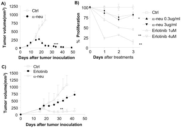
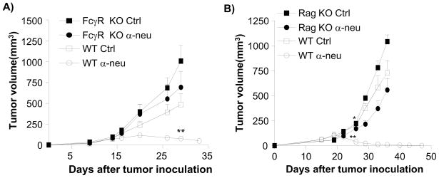
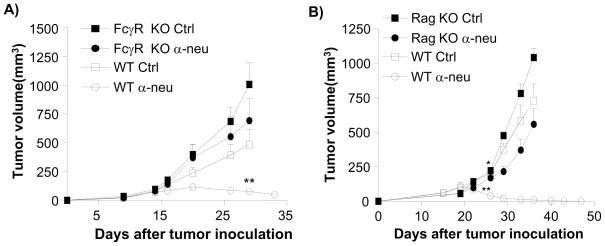
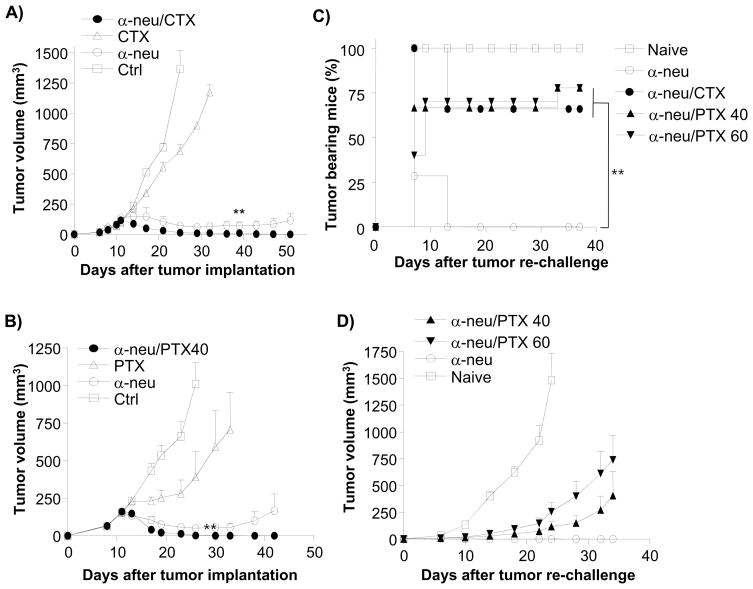
Anti-HER2/neu antibody therapy is reported to mediate tumor regression by interrupting oncogenic signals and/or inducing FcR-mediated cytotoxicity. Here, we demonstrate that the mechanisms of tumor regression by this therapy also require the adaptive immune response. Activation of innate immunity and T cells, initiated by antibody treatment, was necessary. Intriguingly, the addition of chemotherapeutic drugs, although capable of enhancing the reduction of tumor burden, could abrogate antibody-initiated immunity leading to decreased resistance to rechallenge or earlier relapse. Increased influx of both innate and adaptive immune cells into the tumor microenvironment by a selected immunotherapy further enhanced subsequent antibody-induced immunity, leading to increased tumor eradication and resistance to rechallenge. This study proposes a model and strategy for anti-HER2/neu antibody-mediated tumor clearance.


Conflict of interest statement

Statement

The authors have declared that no conflict of interest exists.

Figures

Figure 1
Figure 1. Anti-neu antibody has limited effect in vitro but has strong effect against tumor in vivo
A) WT BALB/c mice (n=5/group) were injected s.c. with 5 × 10⁵ TUBO cells and treated with 100 μg of anti-neu (α-neu) or isotype control (Ctrl) antibody on days 14 and 21. The tumor growth was measured and compared twice a week. ***, p < 0.005 compared with isotype control group after day 23. One of five representative experiments is shown. B) TUBO cells (1 × 10⁵ cells/well) were plated in a monolayer and incubated with Erlotinib (1–4 μmol/L) or anti-neu antibody (0.3–3 μg/mL). Control groups received isotype-control antibody (Ctrl). Relative proliferation, reflected by metabolic activity, was evaluated at indicated times by MTT assay and graphed as percent of isotype control. Mean±SD; *, p < 0.05; **, p < 0.005 compared with isotype control. One of two representative experiments is shown. C) TUBO bearing BALB/c mice (n=5/group) were treated with four times with 100 μg of anti-neu antibody (α-neu) every other day and with 500 μg of Erlotinib every day for 7 days from day 18. *, p < 0.05 compared with the control group from day 33; **, p < 0.01 compared with erlotinib-treated group after day 26. One of two representative experiments is shown.
Figure 2
Figure 2. Anti-tumor effect of antibody depends on both FcR and adaptive immune system
A) TUBO-bearing Fcγ receptor KO and WT BALB/c mice (n=6–8/group) were treated with 100 μg of anti-neu or isotype control antibody on days 14 and 21. **, p < 0.01 compared with WT isotype control group after day 29. One of three experiments is shown. B) TUBO-bearing Wt and Rag-1−/− mice (n=5–7/group) were treated with 100 μg of anti-neu antibody (α-neu) or isotype control (Ctrl) on days 18 and 25. *, p < 0.05; **, p < 0.005 compared to isotype control groups of each mouse strain as A. One of three experiments is shown. Increase of lymphocytes inside tumor is seen in Figure S1.
Figure 3
Figure 3. The therapeutic effect of anti-neu antibody treatment requires CD8+ cells and induces memory T cell responses
A) WT BALB/c mice (n=5–10/group) were injected s.c. with 5 × 10⁵ TUBO and treated with 100 μg of anti-neu antibody (α-neu) on days 10, 17, and 24. CD8-depleting antibody (YTS169.4.2, 200μg/mouse) was administered every 3 days, starting on day 9. *, p < 0.05; **, p < 0.005 compared to anti-neu antibody-treated WT mice. One of three experiments is shown. B) Neu Tg F1 mice (n=6/group) were injected with 3 × 10⁵ TUBO cells and treated with 100 μg of anti-neu antibody (α-neu) on days 11 and 18. CD8-dpeleting antibody (YTS169.4.2, 200μg/mouse) was administered on the same days. *, p < 0.05 compared to anti-neu antibody-treated group. One of two experiments is shown. The data from other CD8-depleting antibodies is shown in Figure S2. C) Tumor-free, antibody-treated BALB/c mice (n=14 pooled from two experiments) were re-challanged s.c. with 5 × 10⁶ TUBO cells on different site from primary tumor at least 1 month after complete rejection of primary tumors. All of mice rejected the secondary tumor. One of two experiments is shown. Anti-neu antibody therapy increases IFNγ + cells in WT (D) or Tg mice (E). D) TUBO bearing mice were treated twice with 150ug of either anti-neu (n=3) or mIgG (n=3) on days 11 and 18. Mice were sacrificed 12 days after the final treatment and splecnoytes were isolated for ELISPOT analysis as described in the materials and methods. ***p<0.0001. E. Splenocytes from neu Tg F1 mice (N=3–5) treated with anti-neu or isotype control antibody were stimulated with 3T3/KB, 3T3/NKB, or TUBO cells. The ratio of splenocytes to APC was 10:1. IFN-γ–producing cells were enumerated by ELISPOT assay. Results were expressed as number of spots per 106 splenocytes. *, p< 0.05; **, p< 0.005 compared with isotype control group. One of three experiments is shown for D and E. Increase of tumor infiltrated lymphocytes was also seen in human samples after antibody treatment (see Figure S2C)
Figure 4
Figure 4. The therapeutic effect of anti-neu antibody depends on endogenous danger signals
A) WT and Myd88−/− BALB/c mice (n=5–7/group) were injected s.c. with 4 × 10⁵ TUBO cells and treated with 100 μg of anti-neu (α-neu) or isotype control (Ctrl) antibody on days 21 and 28. *, p<0.01 compared to anti-neu antibody treated Myd88−/− mice. One of two experiments is shown. B) TUBO-bearing WT BALB/c mice (n=4/group) were treated with 100 μg of anti-neu antibody (α-neu) and 100 μg of neutralizing anti-HMGB-1 antibody on days 14 and 21. **, p < 0.005, compared to anti-HMGB-1 antibody-treated group. One of four experiments is shown.
Figure 5
Figure 5. Administration of chemotherapeutics after antibody treatment enhances primary tumor reduction but reduces immunity induced by anti-neu antibody
WT BALB/c mice (n=5–10/group) were injected s.c. with 5 × 10⁵ TUBO cells and treated with 100 μg of anti-neu antibody (α-neu) on days 11 and 16. Select chemotherapeutic agents were injected i.p. at different time points. One of three experiments is shown. (A) 100 mg/kg of Cyclophosphamide (CTX) was injected i.p. on days 16, 23, and 33. (B) 40 mg/kg of Paclitaxel (PTX) weas injected i.p. on days 14 and 19. Treated, tumor-free mice were re-challenged with 2 × 10⁶ TUBO cells when primary tumor was not detected for at least 30 days. Percent tumor-bearing mice (C) and mean tumor volume (D) are shown. Reduced T cell proliferation was seen in Figure S3.

Comment in

  • Her 2 in 1.
    Smyth MJ, Stagg J. Smyth MJ, et al. Cancer Cell. 2010 Aug 9;18(2):101-2. doi: 10.1016/j.ccr.2010.07.009. Cancer Cell. 2010. PMID: 20708150

References

    1. Aomatsu T, Komatsu H, Yoden A, Hosomi A, Miyazaki H, Sogo T, Inui A, Fujisawa T, Tamai H. Fulminant hepatitis B and acute hepatitis B due to intrafamilial transmission of HBV after chemotherapy for non-Hodgkin's lymphoma in an HBV carrier. Eur J Pediatr. 2010;169:167–171. - PubMed
    1. Apetoh L, Ghiringhelli F, Tesniere A, Obeid M, Ortiz C, Criollo A, Mignot G, Maiuri MC, Ullrich E, Saulnier P, et al. Toll-like receptor 4-dependent contribution of the immune system to anticancer chemotherapy and radiotherapy. Nat Med. 2007;13:1050–1059. - PubMed
    1. Arnould L, Gelly M, Penault-Llorca F, Benoit L, Bonnetain F, Migeon C, Cabaret V, Fermeaux V, Bertheau P, Garnier J, et al. Trastuzumab-based treatment of HER2-positive breast cancer: an antibody-dependent cellular cytotoxicity mechanism? Br J Cancer. 2006;94:259–267. - PMC - PubMed
    1. Burgdorf S, Scholz C, Kautz A, Tampe R, Kurts C. Spatial and mechanistic separation of cross-presentation and endogenous antigen presentation. Nat Immunol. 2008;9:558–566. - PubMed
    1. Chen GY, Tang J, Zheng P, Liu Y. CD24 and Siglec-10 selectively repress tissue damage-induced immune responses. Science. 2009;323:1722–1725. - PMC - PubMed

Publication types