. 2010 Oct 25;406(2):312-21.
doi: 10.1016/j.virol.2010.07.021. Epub 2010 Aug 13.

BST-2 mediated restriction of simian-human immunodeficiency virus

Affiliations

BST-2 mediated restriction of simian-human immunodeficiency virus

Autumn Ruiz et al. Virology. .

Abstract

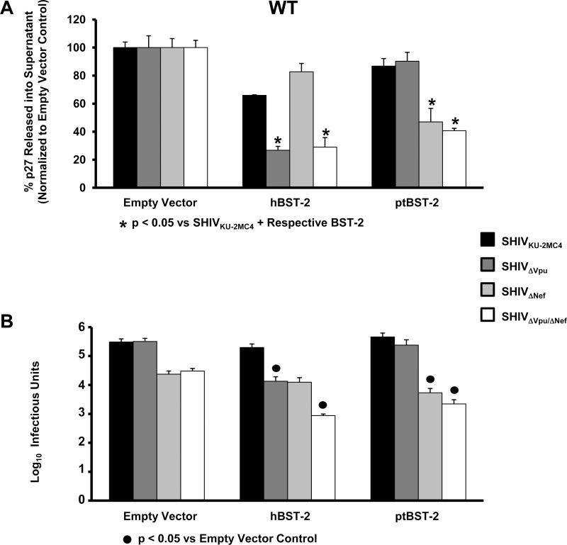
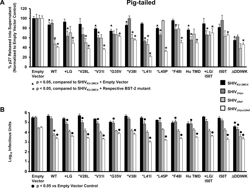
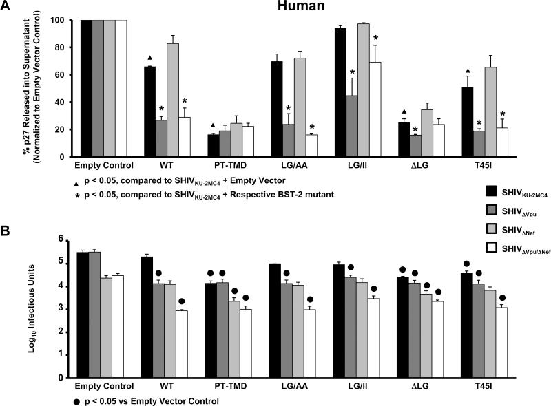
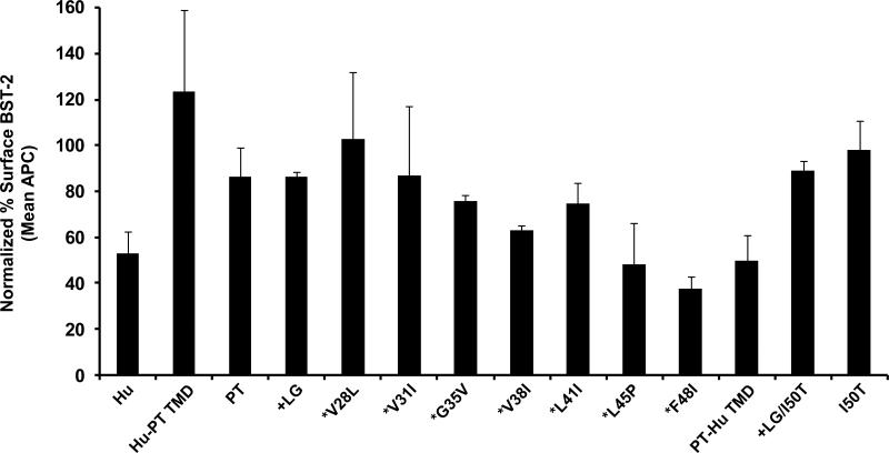
Pathogenic simian-human immunodeficiency viruses (SHIV) contain HIV-1 Vpu and SIV Nef, both shown to counteract BST-2 (HM1.24; CD317; tetherin) inhibition of virus release in a species-specific manner. We show that human and pig-tailed BST-2 (ptBST-2) restrict SHIV. We found that sequential "humanization" of the transmembrane domain (TMD) of the pig-tailed BST-2 (ptBST-2) protein resulted in a fluctuation in sensitivity to HIV-1 Vpu. Our results also show that the length of the TMD in human and ptBST-2 proteins is important for BST-2 restriction and susceptibility to Vpu. Taken together, our results emphasize the importance of tertiary structure in BST-2 antagonism and suggests that the HIV-1 Vpu transmembrane domain may have additional functions in vivo unrelated to BST-2 antagonism.


Figures

Figure 1
Figure 1
Sequence analysis of BST-2 genes amplified from rhesus (A) and pig-tailed (B) macaques. RNA was isolated from either spleen tissue or PBMC from five rhesus macaques and seven pig-tailed macaques. The bst-2 gene was amplified from each of these samples and bulk sequenced. The sequences obtained from the rhesus macaques are presented along with the sequences of rhBST-2 from the genomic database. The BST-2 proteins used in this study for both rhesus and pig-tailed macaques were used as references and the dashes represent similar identity.
Figure 1
Figure 1
Sequence analysis of BST-2 genes amplified from rhesus (A) and pig-tailed (B) macaques. RNA was isolated from either spleen tissue or PBMC from five rhesus macaques and seven pig-tailed macaques. The bst-2 gene was amplified from each of these samples and bulk sequenced. The sequences obtained from the rhesus macaques are presented along with the sequences of rhBST-2 from the genomic database. The BST-2 proteins used in this study for both rhesus and pig-tailed macaques were used as references and the dashes represent similar identity.
Figure 2
Figure 2
The amino acid sequence of the N-terminal region of human and pig-tailed BST-2 proteins, and the BST-2 mutants analyzed in this study.
Figure 3
Figure 3
Expression of parental and mutant BST-2 proteins analyzed in this study. 293 cells were transfected with vectors expressing each of the BST-2 proteins. At 48 hours, cell lysates were collected and the nuclei removed through centrifugation. The lysates were boiled in sample reducing buffer and protein expression was examined through Western blot analysis using a rabbit polyclonal anti-BST-2 antibody (NIH). Panel A. 293 cells transfected with vectors expressing human parental and mutant BST-2 proteins. Panel B. 293 cells transfected with vectors expressing pig-tailed parental and mutant BST-2 proteins.
Figure 4
Figure 4
BST-2 dependent down-regulation of SHIV virion release from cells transfected with hBST-2 and ptBST-2. Panel A. p27 release assay. 293 cells were co-transfected with vectors expressing proviral DNA from one of four SHIV (SHIVKU-2MC4, SHIVΔVpu, SHIVΔNef or SHIVΔVpu/ΔNef) and a vector expressing either the hBST-2 or ptBST-2 protein. At 48 hours post-transfection, the supernatants and cell lysates were collected and cleared of cellular debris and nuclei by centrifugation. The p27 content of both the supernatant and cell lysate from each sample was quantified using a p27 antigen capture assay (Zeptometrix) and the percent p27 release calculated. All conditions were run at least three separate times, normalized to their respective SHIV empty vector controls, and the average percent p27 release and standard error calculated. Significance in the restriction of p27 release for the SHIVKU-2MC4 samples was determined with respect to the parental SHIVKU-2MC4 empty vector control using a Student's t-test (▲). Significance in the restriction of p27 release for the SHIVΔVpu, SHIVΔNef, and SHIVΔVpu/ΔNef samples was calculated with respect to the SHIVKU-2MC4 in the presence of each respective BST-2 using a Student's t-test (*). Panel B. Infectious units assay using TZM-bl indicator cells. Supernatants from transfections described above were added to the TZM-bl cells and serially diluted. At 48hrs post-infection, cells were washed, fixed, and stained for 2 hours. The TCID50 for each supernatant was calculated based on wells containing cells expressing β-galactosidase. All conditions were run at least three times and the TCID50 calculated. The average TCID50 and standard error were calculated. Significance in the restriction of infectivity was determined with respect to the parental SHIV empty vector control using a Student's t-test with p<0.05 considered significant (●).
Figure 5
Figure 5
BST-2 dependent down-regulation of SHIV virion release from cells transfected with vectors expressing ptBST-2 mutants. Panel A. p27 release assay. 293 cells were co-transfected with vectors expressing proviral DNA from one of four SHIV (SHIVKU-2MC4, SHIVΔVpu, SHIVΔNef or SHIVΔVpu/ΔNef) and a vector expressing either the hBST-2 or ptBST-2 protein. At 48 hours post-transfection, the supernatants and cell lysates were collected and cleared of cellular debris and nuclei by centrifugation. The p27 content of both the supernatant and cell lysate from each sample was quantified using a p27 antigen capture assay (Zeptometrix) and the percent p27 release calculated. All conditions were run at least three separate times, normalized to their respective SHIV empty vector controls, and the average percent p27 release and standard error calculated. Significance in the restriction of p27 release for the SHIVKU-2MC4 samples was determined with respect to the parental SHIVKU-2MC4 empty vector control using a Student's t-test (▲). Significance in the restriction of p27 release for the SHIVΔVpu, SHIVΔNef, and SHIVΔVpu/ΔNef samples was calculated with respect to the SHIVKU-2MC4 in the presence of each respecitve BST-2 using a Student's t-test (*). Panel B. Infectious units assay using TZM-bl indicator cells. Supernatants from transfections described above were added to the TZM-bl cells and serially diluted. At 48hrs post-infection, cells were washed, fixed, and stained for 2 hours. The TCID50 for each supernatant was calculated based on wells containing cells expressing β-galactosidase. All conditions were run at least three times and the TCID50 calculated. The average TCID50 and standard error were calculated. Significance in the restriction of infectivity was determined with respect to the parental SHIV empty vector control using a Student's t-test with p<0.05 considered significant (●).
Figure 6
Figure 6
BST-2 dependent down-regulation of SHIV virion release from cells transfected with vectors expressing hBST-2 mutants. Panel A. p27 release assay. 293 cells were co-transfected with vectors expressing proviral DNA from one of four SHIV (SHIVKU-2MC4, SHIVΔVpu, SHIVΔNef or SHIVΔVpu/ΔNef) and a vector expressing either the hBST-2 or ptBST-2 protein. At 48 hours post-transfection, the supernatants and cell lysates were collected and cleared of cellular debris and nuclei by centrifugation. The p27 content of both the supernatant and cell lysate from each sample was quantified using a p27 antigen capture assay (Zeptometrix) and the percent p27 release calculated. All conditions were run at least three separate times, normalized to their respective SHIV empty vector controls, and the average percent p27 release and standard error calculated. Significance in the restriction of p27 release for the SHIVKU-2MC4 samples was determined with respect to the parental SHIVKU-2MC4 empty vector control using a Student's t-test (▲). Significance in the restriction of p27 release for the SHIVΔVpu, SHIVΔNef, and SHIVΔVpu/ΔNef samples was calculated with respect to the SHIVKU-2MC4 in the presence of each respective BST-2 using a Student's t-test (*). Panel B. Infectious units assay using TZM-bl indicator cells. Supernatants from transfections described above were added to the TZM-bl cells and serially diluted. At 48hrs post-infection, cells were washed, fixed, and stained for 2 hours. The TCID50 for each supernatant was calculated based on wells containing cells expressing β-galactosidase. All conditions were run at least three times and the TCID50 calculated. The average TCID50 and standard error were calculated. Significance in the restriction of infectivity was determined with respect to the parental SHIV empty vector control using a Student's t-test with p<0.05 considered significant (●).
Figure 7
Figure 7
The down-regulation of cell surface expression of ptBST-2 mutants by Vpu. To measure surface down-regulation of BST-2 mutants in the presence of HIV-1 Vpu, 293 cells were seeded on a 12-well format and transfected using Lipofectamine 2000. For each transfection, 0.25 μg of pCG-GFP, 0.5 μg of pVphu or mock DNA plasmid, and 0.06 μg of each BST-2 noted above were used. At 24 hours, the cells were reacted with a mouse anti-HM1.24 antibody, then with an APC-conjugated goat anti-mouse antibody, followed by fixation. The cells were analyzed by two-color flow cytometry. For each BST-2 mutant, the APC mean fluorescent intensity (MFI) of high GFP expressing cells in the presence of Vphu was normalized to the MFI of cells without Vphu. The data are represented as percent BST-2 remaining on the surface of transfected cells in the presence of Vphu. All conditions were run at least twice and the standard deviation calculated.

Similar articles

Cited by

References

    1. Aiken C, Trono D. Nef stimulates human immunodeficiency virus type 1 proviral DNA synthesis. J Virol. 1995;69(8):5048–5056. - PMC - PubMed
    1. Ali MS, Hammonds J, Ding L, Spearman P. CAML does not modulate tetherin-mediated restriction of HIV-1 particle release. PLos One. 2010;5(2):e9005. - PMC - PubMed
    1. Andrew AJ, Miyagi E, Kao S, Strebel K. The formation of cysteine-linked dimers of BST-2/tetherin is important for inhibition of HIV-1 virus release but not for sensitivity to Vpu. Retrovirology. 2009;6:80. - PMC - PubMed
    1. Cao W, Bover L, Cho M, Wen X, Hanabuchi S, Bao M, Rosen DB, Wang YH, Shaw JL, Du Q, Li C, Arai N, Yao Z, Lanier LL, Liu YJ. Regulation of TLR7/9 responses in plasmacytoid dendritic cells by BST2 and ILT7 receptor interaction. J. Exp. Med. 2009;206(7):1603–1604. - PMC - PubMed
    1. Chowers MY, Spina CA, Kwoh NJ, Fitch NJ, Richman DD, Guatelli JC. Optimal infectivity in vitro of human immunodeficiency virus type 1 requires an intact nef gene. J Virol. 1994;68(5):2906–2014. - PMC - PubMed

Publication types

MeSH terms

Substances