. 2010 Dec 15;229(1-2):91-7.
doi: 10.1016/j.jneuroim.2010.07.007. Epub 2010 Aug 12.

Involvement of IFN-γ and perforin, but not Fas/FasL interactions in regulatory T cell-mediated suppression of experimental autoimmune encephalomyelitis

Affiliations

Involvement of IFN-γ and perforin, but not Fas/FasL interactions in regulatory T cell-mediated suppression of experimental autoimmune encephalomyelitis

Tara Beeston et al. J Neuroimmunol. .

Abstract

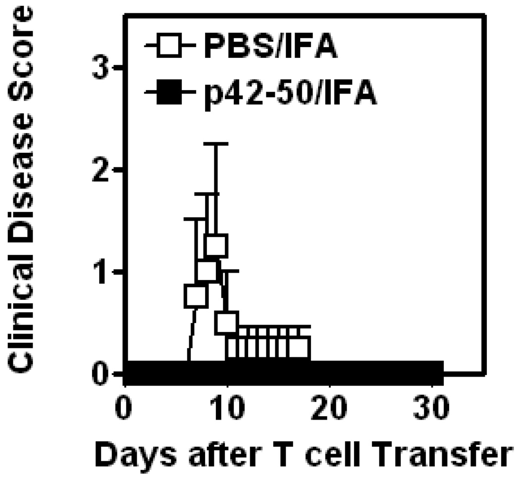
Autoaggressive, myelin-reactive T cells are involved in multiple sclerosis and its prototype experimental autoimmune encephalomyelitis (EAE) in mice. A peripheral negative feedback mechanism involving regulatory CD4+ and CD8+T cells (Treg) operates to suppress disease-mediating T cell responses. We have recently characterized a novel population of Qa-1a-restricted, TCR-peptide-reactive CD8αα+TCRαβ+ Treg that induce apoptotic depletion of the encephalitogenic Vβ8.2 cells in vivo and provide protection from EAE. Here we have used mice deficient in perforin, Fas/FasL and IFN-γ molecules to investigate their role in Treg-mediated regulation of EAE. Data show that Fas/FasL interactions are not involved, but regulation mediated by Treg is dependent on the presence of IFN-γ and the perforin pathway. These data provide a molecular mechanism of Treg-mediated killing of the pathogenic T cells and have important implications in the design of immune interventions for demyelinating disease.


Figures

Figure 1
Figure 1. Vaccination with p42–50 protects B10.PL.lpr mice from active EAE
Groups (4 mice in each) of wildtype B10.PL., B10.PL.lpr and B10.PL.gld mice were vaccinated i.p with 10 µg p42–50/IFA or PBS/IFA. 7 days later EAE was actively induced following immunization with MBPAc1–9/CFA/PTx. Mice were monitored daily for signs of clinical disease, and mean disease score (+/− SEM) of each group is shown. Data are representative of 3 independent experiments.
Figure 2
Figure 2. Vaccination with p42–50 protects B10.PL.gld mice from passive EAE
Groups (4 mice in each) of B10.PL.gld mice were vaccinated i.p with 10 µg p42–50/IFA or PBS/IFA. 7 days later, mice received 1.85×10⁶ MBP-reactive CD4+ T cells i.v. Mice were monitored daily for signs of clinical EAE disease. Mean score (+/− SEM) of each group is shown. Data are representative of two independent experiments.
Figure 3
Figure 3. The anti-MBP T cell response is suppressed in mice vaccinated with p42–50
Groups (4–5 mice in each) of mice were vaccinated i.p with 10 µg p42–50. 8 days later mice were injected with 100 µg MBPAc1–20/CFA/PTx. 10 days later DLN were harvested, and proliferation (upper panel) and IFN-γ secretion (lower panel) in response to an in vitro recall at an optimal concentration of 10 µg/ml MBPAc1–20 was analyzed. Data are representative of two independent experiments.
Figure 4
Figure 4. Successful TCR-peptide vaccination requires a Th1 environment
Groups (6–8 mice in each) of mice were nasally instilled with 10 µg B5 either without cytokines or with IL-4 (10 pg/mouse) or IL-12 (0.3 µg/mouse) to deviate the response towards a Th2 or Th1 direction, respectively (top panel). Active EAE was induced five days later with MBP/CFA/PTx. Mean disease score of each group is shown. IFN-γ is required for p42–50 to protect against EAE (lower panel). Groups (6–8 mice in each) of B10.PL IFN-γ knockout mice were vaccinated with PBS/IFA or p42–50/IFA as indicated. 7 days later mice were immunized with 200 µg MBPAc1–9/CFA/PTx for the active induction of EAE. Mean clinical disease score is shown.
Figure 5
Figure 5. Vaccination with p42–50 in a Th2-like environment potentiates EAE in B10.PL.wt and B10.PL.lpr mice
Groups (6–8 mice in each) of mice were vaccinated with 10 µg p42–50 in the presence of IL-4 (10 pg/mouse) to create a Th2 environment. Seven days later EAE was actively induced by MBPAc1–9/CFA/PTx immunization. Mean disease score of each group is shown.

References

    1. Steinman L. Multiple sclerosis: a coordinated immunological attack against myelin in the central nervous system. Cell. 1996;85:299. - PubMed
    1. Martin R. HLA class I: friend and foe of multiple sclerosis. Nat Med. 2008;14:1150. - PubMed
    1. Traugott U, Reinherz EL, Raine CS. Multiple sclerosis: distribution of T cell subsets within active chronic lesions. Science. 1983;219:308. - PubMed
    1. Hauser SL, Bhan AK, Gilles F, Kemp M, Kerr C, Weiner HL. Immunohistochemical analysis of the cellular infiltrate in multiple sclerosis lesions. Ann. Neurol. 1986;19:578. - PubMed
    1. Huseby ES, Liggitt D, Brabb T, Schnabel B, Ohlen C, Goverman J. A pathogenic role for myelin-specific CD8(+) T cells in a model for multiple sclerosis. J Exp. Med. 2001;194:669. - PMC - PubMed

Publication types

MeSH terms

LinkOut - more resources