Review
. 2010 Sep;35(9):514-21.
doi: 10.1016/j.tibs.2010.03.005. Epub 2010 Aug 12.

The 3D structures of VDAC represent a native conformation

Affiliations
Review

The 3D structures of VDAC represent a native conformation

Sebastian Hiller et al. Trends Biochem Sci. 2010 Sep.

Abstract

The most abundant protein of the mitochondrial outer membrane is the voltage-dependent anion channel (VDAC), which facilitates the exchange of ions and molecules between mitochondria and cytosol and is regulated by interactions with other proteins and small molecules. VDAC has been studied extensively for more than three decades, and last year three independent investigations revealed a structure of VDAC-1 exhibiting 19 transmembrane beta-strands, constituting a unique structural class of beta-barrel membrane proteins. Here, we provide a historical perspective on VDAC research and give an overview of the experimental design used to obtain these structures. Furthermore, we validate the protein refolding approach and summarize the biochemical and biophysical evidence that links the 19-stranded structure to the native form of VDAC.


Figures

Figure 1
Figure 1. Experimental approaches for VDAC characterization
The two primary production sources of VDAC protein are indicated by gray rectangles (native expression and inclusion bodies). Black rectangles designate selected preparative states of VDAC together with characterization techniques that have been used in these states (AFM: atomic force microscopy; EM: electron microscopy; NMR: nuclear magnetic resonance; VG: voltage gating; X-ray: X-ray crystallography). The small figures illustrate the experimental findings, reproduced with permission from [4,11,13,22,48,49][SC4]. The black arrows designate the preparative paths leading to individual experimental states and the procedures along these paths are given in magenta. Blue arrows highlight consistent experimental findings from the same technique applied to protein samples prepared by native expression in mitochondria and heterologous expression in inclusion bodies. All figures were prepared using PYMOL [50].
Figure 2
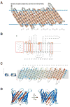
Figure 2. Predicted and experimentally determined topologies of VDAC
(A) Structural model of VDAC by Forte et al. as determined by the Delphi algorithm in 1987 (reproduced with permission from [16]). Bold letters indicate amino acid residues lining the aqueous pore, small letters interact with the hydrophobic membrane. The 19 β-strands as observed in the 3D X-ray and NMR structure are indicated by red rectangles. (B) Model developed by the Colombini and coworkers, suggesting the VDAC pore is formed by 13 β-strands and 1 α-helix (reproduced with permission from [26]). Residues in black circles denote the edges of the β-strands. Residues marked red were proposed by Colombini and coworkers to comprise the voltage sensor [37]. (C) Topology of VDAC-1 with 19 β-strands as determined by 3D NMR and X-ray structures [21-23]. Bold residues point towards the hydrophobic phase, the other residues into the aqueous pore. The blue cylinders indicate the alpha helix which extends into the pore but is not part of the wall. (D) 3D structure of VDAC-1 (blue; PDB-entry: 3EMN). The voltage sensor proposed by Colombini and coworkers is indicated in red (compare also to panel B) [37].
Figure 3
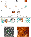
Figure 3. Structural features of VDAC
(A) Examples of data from each of the three experimental approaches for the VDAC-1 structure determination, from left to right: [21-23]. (B) Resulting 3D structures of VDAC. (C) Overlay of the three structures in top and side view, yielding an RMSD of 1.5 Å over 171 Cα atoms between the NMR-only and X-ray only structure. (D–G) Structural features of VDAC. (D, E) Cartoon representation of VDAC-1 viewed parallel (D) and perpendicular (E) to the plane of the membrane. The VDAC-1 protein structure is colored from the N-terminus in blue to the C-terminus in red. VDAC forms a large pore conducive to high metabolite flux representing the open conformation. The N-terminal helix contacts the inner barrel wall and is not part of the pore. (F) The unique parallel strand arrangement between strand 1 and 19 revealing a new class of β-barrel proteins. (G) Location of Glu 73 residing in the lipidic environment. (H) The 3D structure overlaid onto EM [4] (left panel) and AFM images [49] (right panel). The arrangement of a VDAC hexamer is putative, based on the observed 2D crystal packing of VDAC-1. In the AFM images the hexamer overlap is poor because of a tilt of the AFM image.
Figure 4
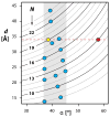
Figure 4
Geometrical constraints for β-barrel membrane proteins. The wall-to-wall diameter d of ideal, circular β-barrels is plotted versus the tilt angle α of the β-strands against the membrane normal, following the approach by Schulz [51]. For a given number of strands, N, the accessible d/α values are located on hyperbolic curves. A 19-stranded barrel with a shear number of 20, corresponding to VDAC-1, is indicated by a yellow circle. Blue circles denote all known monotopic integral β-barrel membrane protein structures. The range 37°–45° of the tilt angles α, which all known such structures are limited to is underlined grey. A hypothetical 13-stranded barrel with a diameter of 34 Å is shown red, resulting in a tilt angle of 58°.

References

    1. Parsons DF, et al. Characteristics of isolated and purified preparations of outer and inner membranes of mitochondria. Ann NY Acad Sci. 1966;137:643–666. - PubMed
    1. Schein SJ, et al. Reconstitution in planar lipid bilayers of a voltage-dependent anion-selective channel obtained from Paramecium mitochondria. J Membr Biol. 1976;30:99–120. - PubMed
    1. Mannella CA, Colombini M. Evidence that the crystalline arrays in the outer membrane of Neurospora mitochondria are composed of the voltage-dependent channel protein. Biochim Biophys Acta. 1984;774:206–214. - PubMed
    1. Guo XW, et al. Molecular design of the voltage-dependent, anion-selective channel in the mitochondrial outer membrane. J Struct Biol. 1995;114:41–59. - PubMed
    1. Colombini M. A candidate for the permeability pathway of the outer mitochondrial membrane. Nature. 1979;279:643–645. - PubMed

Publication types

MeSH terms

Substances

LinkOut - more resources