. 2010 Aug 17;19(2):220-31.
doi: 10.1016/j.devcel.2010.07.007.

beta-Catenin primes organizer gene expression by recruiting a histone H3 arginine 8 methyltransferase, Prmt2

Affiliations

beta-Catenin primes organizer gene expression by recruiting a histone H3 arginine 8 methyltransferase, Prmt2

Shelby A Blythe et al. Dev Cell. .

Abstract

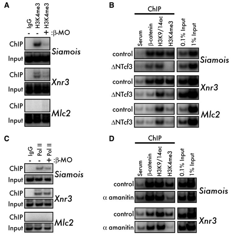
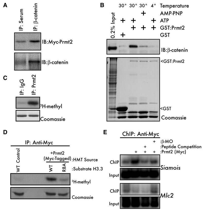
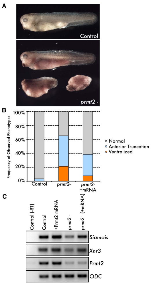
An emerging concept in development is that transcriptional poising presets patterns of gene expression in a manner that reflects a cell's developmental potential. However, it is not known how certain loci are specified in the embryo to establish poised chromatin architecture as the developmental program unfolds. We find that, in the context of transcriptional quiescence prior to the midblastula transition in Xenopus, dorsal specification by the Wnt/beta-catenin pathway is temporally uncoupled from the onset of dorsal target gene expression, and that beta-catenin establishes poised chromatin architecture at target promoters. beta-catenin recruits the arginine methyltransferase Prmt2 to target promoters, thereby establishing asymmetrically dimethylated H3 arginine 8 (R8). Recruitment of Prmt2 to beta-catenin target genes is necessary and sufficient to establish the dorsal developmental program, indicating that Prmt2-mediated histone H3(R8) methylation plays a critical role downstream of beta-catenin in establishing poised chromatin architecture and marking key organizer genes for later expression.


Figures

Figure 1
Figure 1. β-catenin target genes are poised for expression before the MBT
(A) Onset of expression of maternal β-catenin target genes (Siamois, Xnr3, Xnr5, and Xnr6). Maternally expressed Ornithine decarboxylase (Odc) is shown as a control for loading. Odc (-RT) indicates no reverse transcriptase, as a control for genomic DNA contamination. (B) Embryos were injected at the 2-cell stage with the β-catenin morpholino (ii-iv) and subsequently at the 4-cell stage (2 dorsal blastomeres) with 2pg siamois (iii) or 300pg β-catenin (iv) mRNAs. The frequency of each representative phenotype is indicated. (C) Embryos were injected into two dorsal blastomeres at the 4-cell stage with 500pg ΔNTcf3-GR mRNA. Wnt/β-catenin activity was inhibited by addition of dexamethasone (Dex) to the culture medium at the indicated stages. Siamois, Xnr3, and ODC mRNA expression was measured by RT-PCR at stage 10. (D) Left panel: occupancy of initiating (CTD pSer5) RNA Pol II in the promoter-proximal regions of the Siamois and Xnr3 loci before (1000-cell stage) or after (Stage 9) the onset of expression as measured by ChIP-QPCR. Right panel: elongating (CTD pSer2) RNA Pol II associated with the 3’ CDS of the same panel of genes at the same timepoints. Binding of RNA Pol II at Xnr6, which is expressed at both stages, is a positive control. Pooled data from three independent experiments are presented as a percentage of input chromatin to facilitate comparison between the 1000-cell stage and Stage 9. The average signal (0.02% input) from a negative control (IgG) ChIP is marked as a dotted line. Error bars are S.E.M. (E) PreMBT occupancy of maternal β-catenin target promoters (Siamois, Xnr3, and Xnr5) or a zygotic β-catenin target promoter (Myf5) by either H3K9/14ac or β-catenin was observed by ChIP on 1000-cell stage embryos. Myosin light chain 2 (Mlc2): negative control locus. “Input” indicates chromatin prior to ChIP (1%). (F) Promoter occupancy by β-catenin or H3K4me3 measured by ChIP on 1000-cell embryos. See also Figure S1.
Figure 2
Figure 2. β-catenin and RNA Pol II establish H3K4me3 at poised promoters
(A) ChIP for H3K4me3 in 1000-cell control and β-catenin depleted (β-MO) embryos. All sibling β-MO embryos were completely ventralized at later stages (not shown). (B) H3K4me3, but not H3K9/14ac, requires β-catenin function. MBT-stage control (upper panel for each gene) and ΔNTcf3 mRNA-injected (500pg, lower panels) embryos were subjected to ChIP for β-catenin, H3K9/14ac, and H3K4me3. (C) 1000-cell stage control and β-catenin depleted (β-MO) embryos were subjected to ChIP for RNA Pol II (CTD pSer5). All sibling β-MO embryos were completely ventralized at later stages (not shown). (D) MBT-stage control (upper panels) and α-amanitin injected (10ng, lower panels) embryos were subjected to ChIP for β-catenin, H3K9/14ac, and H3K4me3.
Figure 3
Figure 3. β-catenin associates with a Histone H3(R8) methyltransferase before the MBT
(A) β-catenin was immunoprecipitated from 16-cell embryos and HMT activity was visualized by fluorography (1 day exposure) for incorporation of [3H] methyl groups into calf thymus histones (top panel). Equal loading of histones is shown by coomassie staining (lower panel). “Input” represents HMT activity in embryo lysates, with activity toward both H3 and H4. (B) β-catenin IP/HMT assays were performed on either wild type (WT) recombinant H3.3 or H3.3 with the indicated point mutations. (C) β-catenin IP/HMT assays were performed on peptides corresponding to unmodified H3 (aa 1-15, lanes 1&2), asymmetrically dimethylated R8 (aa 1-15, lane 3), unmodified H3 (aa 1-21, lanes 4&5), acetylated K9 (aa 1-20, lane 6) and trimethylated K9 (aa 1-24, lane 7). (D) MBT-stage control and lithium chloride treated (LiCl, 300mM for 10 minutes, 1 hour prior to harvest) embryos were subjected to ChIP for either H3K9me1 or H3K9me3. (E) ChIP was performed as described in panel D, using instead antibodies to either H3R8me2a or H3R8me2s (Pal et al., 2004). (F) ChIP was performed on 1000-cell stage control and β-catenin knockdown (β-MO) embryos with the Active Motif H3R8me2a antiserum. See also Figure S2.
Figure 4
Figure 4. β-catenin interacts with the Histone H3(R8) methyltransferase Prmt2
(A) Myc-tagged mouse Prmt2 (500pg) was expressed in Xenopus embryos and embryo lysates (early blastula) were immunoprecipitated with anti-β-catenin or pre-immune serum and subjected to western blot with either anti-myc (upper panel) or anti-β-catenin antibodies. (B) PreMBT embryo lysates were incubated for 1 hour with GST-Prmt2 beads at 4° or 30°C and with ATP or the nonhydrolyzable ATP analog AMP-PNP. Bound proteins were eluted and β-catenin was visualized by western blot (upper panel). GST beads alone were used as a negative control and the relative amounts of bait proteins in each lane were visualized by coomassie staining (lower panel, arrowheads). “Input” indicates non-fractionated input embryo lysate. (C) Endogenous Prmt2 from mouse embryonic stem cells was subjected to an IP/HMT assay using recombinant H3.3 as a substrate. Rabbit IgG was the negative control. (D) Activity of Myc-Prmt2 from 32- to 128-cell Xenopus embryos was measured by IP/HMT assay using wild-type (WT) or R8A histone H3.3 as substrates. Non-injected embryos served as negative controls. (E) Wild type or β-MO injected 1000-cell embryos expressing Myc-Prmt2 were subjected to ChIP using the anti-Myc-tag antibody. Non-injected embryos served as a negative control. The specificity of ChIP was confirmed by including a 200-fold excess of the Myc-peptide (lane 3) in the IP.
Figure 5
Figure 5. Maternal Prmt2 is necessary for dorsal specification
(A and B) Transplantation and fertilization of maternal Prmt2-depleted (prmt2-) oocytes results in a range of ventralized tadpole-stage phenotypes (A, lower panel) compared to controls, which develop normally (A, top panel). The mean frequency of phenotypes arising from maternal Prmt2 depletion is plotted in B (see text for details). The frequency of ventralized embryos (both partial and complete) is reduced by co-injection of Prmt2 mRNA (“prmt2-/+mRNA”). (C) Blastula stage (stage 9) siamois and xnr3 expression was measured in prmt2- and rescued (“prmt2- (+mRNA”)) host transfer embryos as compared to control (non-depleted) and Prmt2 mRNA injected embryos. As a control for the efficiency of knockdown, prmt2 was measured. Note that the rescuing mRNA (1ng of mouse Prmt2, injected into oocytes) is not amplified by the Xenopus prmt2 primers used here. Expression of rescuing mRNA was confirmed by western blot (not shown). Embryos expressing Prmt2 mRNA alone developed identically to controls, with no dorsoventral defects (not shown). See also Figure S3.
Figure 6
Figure 6. Directing Prmt2 to β-catenin target gene promoters is sufficient to drive dorsal specification
(A) Schematic of the Prmt2:ΔNLef1 chimeric construct. To direct Prmt2 to Tcf/Lef DNA binding sites, the DNA-binding HMG domain of mouse Lef1 was fused to the C-terminus of mouse Myc-Prmt2. (B) 4-cell embryos were injected with either wild-type (WT) or catalytically inactive SAM-binding mutant G159,161R (GG) Prmt2:ΔNLef1 into two ventral blastomeres. Phenotypes (left panel) were scored at late-neurula / early tailbud stages. Mean frequency of secondary axis formation (both fully and partially extended) resulting from expression of wild-type or GG mutant Prmt2:ΔNLef1 is plotted on the right. N=197, 172, and 81 embryos for control, WT, and GG mutant, respectively. Error bars are SEM. P=0.017 (two-tailed Student’s T-Test) for the 4 independent trials where WT and mutant were compared directly. Equal expression of wild type and GG mutant Prmt2:ΔNLef1 was verified by western blot for the myc tag (inset). (C) Embryos were depleted for β-catenin (β-MO, panels ii-iv) and subsequently injected with 500pg of either Prmt2:ΔNLef1 (iii) or Prmt5:ΔNLef1 (iv) mRNA. Rescue of β-MO-induced ventralization (ii) was measured at tadpole stages. Note the rescue of the anterior-most, dorsally derived cement gland and eye in panel iii, compared to control, non-injected embryos (i). The percentages in the upper right corner of each panel indicate the frequency at which the phenotypes shown were observed. (D) Embryos were depleted for β-catenin (β-MO) and subsequently injected with Prmt2:ΔNLef1 or ΔNLef1 mRNA as in (C). Expression of Siamois and Xnr3 was measured by RT-PCR at stage 10. EF1α expression is shown as a loading control. (E) Prmt2:ΔNLef1 and Carm1:ΔNLef1 mRNAs (500pg) were injected and RT-PCR was performed as described for C and D.

Comment in

Similar articles

Cited by

References

    1. Akkers RC, van Heeringen SJ, Jacobi UG, Janssen-Megens EM, Francoijs KJ, Stunnenberg HG, Veenstra GJ. A hierarchy of H3K4me3 and H3K27me3 acquisition in spatial gene regulation in Xenopus embryos. Dev Cell. 2009;17:425–434. - PMC - PubMed
    1. Baugh LR, Demodena J, Sternberg PW. RNA Pol II Accumulates at Promoters of Growth Genes During Developmental Arrest. Science. 2009;324:92–94. - PubMed
    1. Bedford MT, Clarke SG. Protein Arginine Methylation in Mammals: Who, What, and Why. Molecular Cell. 2009;33:1–13. - PMC - PubMed
    1. Behrens J, von Kries JP, Kühl M, Bruhn L, Wedlich D, Grosschedl R, Birchmeier W. Functional interaction of beta-catenin with the transcription factor LEF-1. Nature. 1996;382:638–642. - PubMed
    1. Belenkaya TY, Han C, Standley HJ, Lin X, Houston DW, Heasman J, Lin X. pygopus Encodes a nuclear protein essential for wingless/Wnt signaling. Development. 2002;129:4089–4101. - PubMed

Publication types

MeSH terms

Associated data