. 2010 Aug 17;19(2):259-69.
doi: 10.1016/j.devcel.2010.07.015.

Focal adhesion kinase is required for intestinal regeneration and tumorigenesis downstream of Wnt/c-Myc signaling

Affiliations

Focal adhesion kinase is required for intestinal regeneration and tumorigenesis downstream of Wnt/c-Myc signaling

Gabrielle H Ashton et al. Dev Cell. .

Erratum in

Abstract

The intestinal epithelium has a remarkable capacity to regenerate after injury and DNA damage. Here, we show that the integrin effector protein Focal Adhesion Kinase (FAK) is dispensable for normal intestinal homeostasis and DNA damage signaling, but is essential for intestinal regeneration following DNA damage. Given Wnt/c-Myc signaling is activated following intestinal regeneration, we investigated the functional importance of FAK following deletion of the Apc tumor suppressor protein within the intestinal epithelium. Following Apc loss, FAK expression increased in a c-Myc-dependent manner. Codeletion of Apc and Fak strongly reduced proliferation normally induced following Apc loss, and this was associated with reduced levels of phospho-Akt and suppression of intestinal tumorigenesis in Apc heterozygous mice. Thus, FAK is required downstream of Wnt Signaling, for Akt/mTOR activation, intestinal regeneration, and tumorigenesis. Importantly, this work suggests that FAK inhibitors may suppress tumorigenesis in patients at high risk of developing colorectal cancer.


Figures

Figure 1
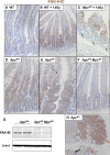
Figure 1. FAK Is Upregulated following Wnt Signaling Activation In Vivo
(A–C) FAK IHC in wild-type (A) and regenerating epithelium (B and C). Note low levels of FAK expression in normal intestinal epithelium (A) which markedly increase in regenerating epithelium (B) 3 days after gamma irradiation. (C) Lack of FAK expression in dying c-Myc-deficient crypts (arrows) 3 days after gamma irradiation. (D and E) FAK IHC in AhCre+Apcfl/fl (labeled Apcfl/fl) intestines 4 days postinduction (PI). IHC shows a clear upregulation of FAK in Apc-deficient intestines (D). This is highlighted at the margin of the Apc-deficient cells where nonrecombined retained villi enterocytes have low levels of FAK protein (black arrow), while Apc-deficient cells (red arrow) have very high levels of FAK protein (E). The reason why there are still wild-type cells retained 4 days following Cre activation is that Apc-deficient cells do not migrate efficiently (see Sansom et al., 2004). (F) FAK IHC in AhCre+Apcfl/flMycfl/fl (labeled Apcfl/flMycfl/fl) intestines 4 days after Apc loss. IHC shows that FAK is not upregulated in double-mutant Apc Myc intestines. (G) Immunoblotting comparing FAK levels in epithelial extracts from AhCre+Apcfl/fl and AhCre+Apcfl/flMycfl/fl intestines 4 days PI. Note there is clearly reduced FAK protein expression in the AhCre+Apcfl/flMycfl/fl intestines. (H) FAK IHC of adenomas of AhCre+Apcfl/+Fak+/+ mice 300 days following Cre induction. Note FAK is upregulated in both adenomas (solid red arrow) and single aberrant crypts (dashed red arrow) compared with normal epithelium (black arrow).
Figure 2
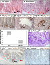
Figure 2. c-Myc Is Required for Intestinal Regeneration
(A) Wild-type regenerating crypts are enlarged following 14 Gy irradiation (black arrows). (B) AhCre+Mycfl/fl intestines day 6 PI, 3 days following 14 Gy irradiation. Note there are now large areas of intestine denuded of crypts with only occasional enlarged regenerative crypts (denoted by black arrow). Dying/cystic crypts are denoted by red arrows. (C) IHC for c-Myc in wild-type intestine day 6 PI. Note c-Myc-positive cells are located at the base of the crypt. (D and E) IHC for c-Myc in AhCre+Mycfl/fl intestine day 6 PI, 3 days following 14 Gy irradiation. The regenerative intestinal crypts (black arrows) are all c-Myc proficient. These are thus defined as escaper crypts. (F) Boxplot showing significant reduction in surviving crypts between AhCre+Mycfl/flRosaMyc/+ (labeled Rosa Myc) and wild-type AhCre+Myc+/+Rosa+/+ mice (Mann-Whitney p = 0.04, n = 4). (G) H&E staining showing absence of functional crypts in AhCre+Mycfl/flRosaMyc/+ (labeled Mycfl/flRosaMyc/+) mice, 72 hr following 14 Gy irradiation. (H) FAK IHC showing FAK expression is generally absent from dying AhCre+Mycfl/fl intestinal crypts 72 hr following irradiation (shown by red arrows). Black arrow denotes regenerating escaper crypt, which has high levels of FAK protein and would express c-Myc (see Figure S1). (I) FAK IHC performed on AhCre+Mycfl/flRosaMyc/+ intestinal epithelium 72 hr following 14 Gy irradiation showing absence of expression in the intestinal crypts.
Figure 3
Figure 3. FAK Is Required for Intestinal Regeneration
(A) H&E staining of small intestine after 14 Gy irradiation showing multiple regenerating crypts in wild-type mice and only one regenerating crypt in AhCre+Fakfl/fl mice (black arrow). (B) Bar graph showing significant decrease in number of regenerating crypts in small intestine of AhCre+Fakfl/fl mice compared with wild-type, 72 hr after 14 Gy irradiation. (C) Corresponding high levels of FAK seen by IHC in FAK-proficient regenerating crypts 72 hr after 14 Gy irradiation. Black arrows show regenerating escaper crypts with high FAK expression. Inset shows close up regenerating crypt with high FAK expression and hence has escaped cre-mediated recombination and not deleted FAK. (D and E) Boxplots showing significantly increased apoptosis and reduced mitosis in AhCre+Fakfl/fl intestine compared with AhCre+Fak+/+ intestine 48 hr following gamma irradiation (Mann-Whitney p > 0.05, n = at least 4). (F) H&E staining of small intestine after 14 Gy irradiation showing multiple regenerating crypts in wild-type mice, and only one regenerating crypt in VillinCreERFakfl/fl mice 72 hr following irradiation (black arrows). (G) Bar graph showing significant decrease in number of regenerating crypts in the small intestine of VillinCreERFakfl/fl mice compared with wild-type, 72 hr after 14 Gy irradiation.
Figure 4
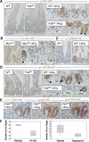
Figure 4. Akt Signaling via FAK Is Required for Efficient Regeneration
(A) IHC for p-AktSer473 shows weak staining in wild-type (first panel) and FAK-deficient crypts (second panel), but increased staining in regenerating crypts following 14 Gy irradiation (third panel upper). No upregulation was observed in FAK-deficient crypts (third panel lower: red arrows); however, in the rare escaper FAK-proficient crypts (black arrows), which efficiently regenerate, high levels of p-Akt were observed. (B) IHC for p-Akt shows that Akt upregulation is c-Myc dependent. First panel shows weak p-Akt staining in nonirradiated c-Myc-deficient intestine. Second panel shows upregulation of p-Akt in a rare escaper c-Myc-proficient regenerating crypt (black arrow), while no discernable p-Akt staining was observed in the dying c-Myc-deficient crypts (red arrows). (C) p-GSK3β (a target of Akt) is activated during regeneration. Left panel, nonir-radiated wild-type crypts showing low levels of p-GSK3β. Right panel shows high levels of p-GSK3β in wild-type regenerating crypts. (D) p-mTOR is upregulated during regeneration but not in FAK-deficient crypts. p-mTOR IHC showing low levels of p-mTOR in wild-type (first panel) and FAK-deficient (second panel) crypts. Following irradiation, increased levels of p-mTOR are observed in wild-type regenerating crypts (third panel upper). In FAK-deficient crypts, p-mTOR is not upregulated (third panel lower: red arrow); however, in the rare escaper regenerating crypts, p-mTOR is upregulated (black arrow). (E) “Back to back” IHC staining showing that the large escaper FAK-proficient regenerative crypts have high levels of p-Akt and p-mTOR expression. (F) Boxplots showing that mice treated with either rapamycin or PI103 have significantly fewer surviving crypts than vehicle treated mice following 14 Gy irradiation (Mann-Whitney, p = > 0.04, n = 4).
Figure 5
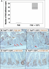
Figure 5. Activation of p-Akt by IGF1 Restores Intestinal Regeneration to FAK-Deficient Intestines
(A) Boxplot showing regenerating FAK-deficient crypts in AhCre+Fakfl/fl intestines treated with IGF1 (Mann-Whitney, p = 0.04), compared with vehicle treated AhCre+Fakfl/fl intestines. (B and C) FAK IHC showing the presence of enlarged FAK-deficient crypts (red arrows), which are the same size as FAK-proficient crypts (black arrows). (D and E) p-Akt and p-mTOR IHC showing all crypts from AhCre+Fakfl/fl intestines treated with IGF1 now have high levels of both p-Akt (D) and p-mTOR (E).
Figure 6
Figure 6. FAK Is Required for Intestinal Transformation following Apc Loss
(A) H&E-stained sections from AhCre+Apcfl/fl (labeled Apcfl/fl) and AhCre+Apcfl/flFakfl/fl (labeled Apcfl/fl Fakfl/fl) intestines 4 days PI. Note that Apc-deficient crypts look much enlarged. (B) FAK IHC in AhCre+Apcfl/fl and AhCre+Apcfl/flFakfl/fl intestines 4 days after Apc loss. IHC shows that FAK expression is lost in doube-mutant Apc Fak intestines. (C) Bar graph showing reduced BrdU positivity and increased apoptosis in AhCre+Apcfl/flFakfl/fl intestinal enterocytes compared with AhCre+Apcfl/fl mice (Mann-Whitney p = 0.04, n ≥ 4). Errors bars represent SD. (D and E) β-catenin and c-Myc IHC performed upon AhCre+Apc+/+ (labeled WT), AhCre+Apcfl/fl and AhCre+Apcfl/flFakfl/fl intestines 4 days PI. Note both Apc and Apc Fak-deficient intestines have increased levels of nuclear β-catenin and c-Myc compared with WT. Nuclear β-catenin is arrowed in the inset. (F and G) p-Akt and p-mTOR IHC performed upon AhCre+Apcfl/fl and AhCre+Apcfl/flFakfl/fl intestines 4 days PI. Note reduction in staining in the double-mutant Apc Fak intestines. (H) Intestinal tumor-free survival of AhCre+Apcfl/+Fak+/+ (blue line) and AhCre+Apcfl/+Fakfl/fl (green line) mice up to 500 days. No AhCre+Apcfl/+Fakfl/fl mice developed intestinal disease. All the AhCre+Apcfl/+ Fak+/+ mice became ill with intestinal tumors with a median onset of 344 days p ≥0.005 log rank.

Comment in

  • Getting one's Fak straight.
    Evan G. Evan G. Dev Cell. 2010 Aug 17;19(2):185-6. doi: 10.1016/j.devcel.2010.07.023. Dev Cell. 2010. PMID: 20708578 Free PMC article.

References

    1. Agochiya M, Brunton VG, Owens DW, Parkinson EK, Paraskeva C, Keith WN, Frame MC. Increased dosage and amplification of the focal adhesion kinase gene in human cancer cells. Oncogene. 1999;18:5646–5653. - PubMed
    1. Amornphimoltham P, Leelahavanichkul K, Molinolo A, Patel V, Gutkind JS. Inhibition of mammalian target of rapamycin by rapamycin causes the regression of carcinogen-induced skin tumor lesions. Clin. Cancer Res. 2008;14:8094–8101. - PMC - PubMed
    1. Andreu P, Colnot S, Godard C, Gad S, Chafey P, Niwa-Kawakita M, Laurent-Puig P, Kahn A, Robine S, Perret C, et al. Crypt-restricted proliferation and commitment to the Paneth cell lineage following Apc loss in the mouse intestine. Development. 2005;132:1443–1451. - PubMed
    1. Athineos D, Sansom OJ. Myc heterozygosity attenuates the phenotypes of APC deficiency in the small intestine. Oncogene. 2010;29:2585–2590. - PubMed
    1. Bach SP, Renehan AG, Potten CS. Stem cells: the intestinal stem cell as a paradigm. Carcinogenesis. 2000;21:469–476. - PubMed

Publication types

MeSH terms

Substances