. 2010 Aug 17;19(2):296-306.
doi: 10.1016/j.devcel.2010.07.019.

Hemocyte-secreted type IV collagen enhances BMP signaling to guide renal tubule morphogenesis in Drosophila

Affiliations

Hemocyte-secreted type IV collagen enhances BMP signaling to guide renal tubule morphogenesis in Drosophila

Stephanie Bunt et al. Dev Cell. .

Abstract

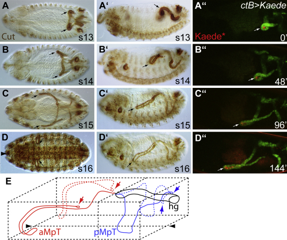
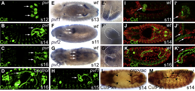
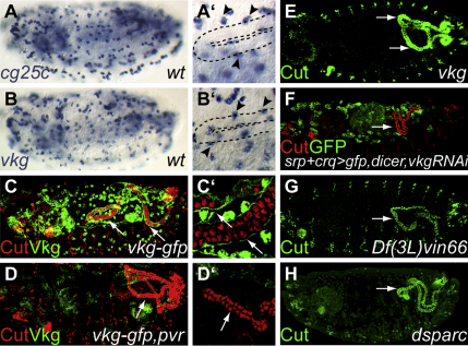
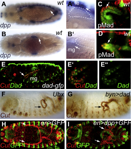
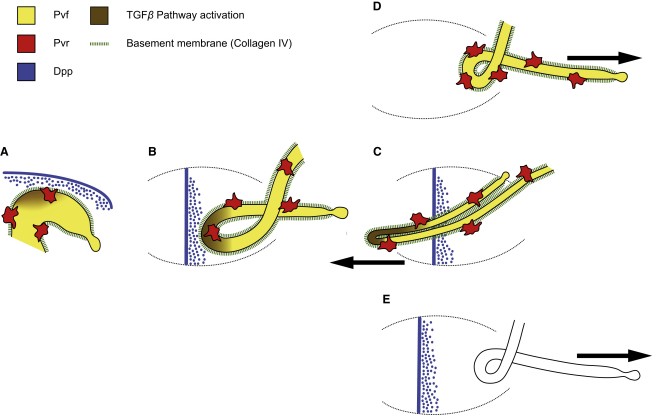
Details of the mechanisms that determine the shape and positioning of organs in the body cavity remain largely obscure. We show that stereotypic positioning of outgrowing Drosophila renal tubules depends on signaling in a subset of tubule cells and results from enhanced sensitivity to guidance signals by targeted matrix deposition. VEGF/PDGF ligands from the tubules attract hemocytes, which secrete components of the basement membrane to ensheath them. Collagen IV sensitizes tubule cells to localized BMP guidance cues. Signaling results in pathway activation in a subset of tubule cells that lead outgrowth through the body cavity. Failure of hemocyte migration, loss of collagen IV, or abrogation of BMP signaling results in tubule misrouting and defective organ shape and positioning. Such regulated interplay between cell-cell and cell-matrix interactions is likely to have wide relevance in organogenesis and congenital disease.


Figures

Figure 1
Figure 1
Malpighian Tubule Morphogenesis Is Highly Reproducible (A–D) Wild-type embryos stained for Cut (brown) to mark the tubules as they elongate, coursing through the body cavity to take up stereotypical positions by stage 16. (A and A′) Stage 13. The four postmitotic tubules start to elongate by cell rearrangement. The anterior tubules bend forming a “kink” region, which projects toward the head (A and A′, arrows). Stage 14 (B and B′) and 15 (C and C′). The anterior tubule kink projects anteriorly on either side of the midgut (B and B′, arrows), and drops ventrally (B′ and C′, arrows). (D and D′) Stage 16. The kink regions of all four tubules lie ventrally, extending toward the midline on either side of the CNS (D and D′, arrows, anterior; midline, large arrowhead in D). (A″–D″) WT embryos expressing the photoconvertible fluorophore Kaede in the tubules (green). Kaede was activated in the kink region at time 0 (Keade, red). Snapshots of a living embryo at times indicated reveal labeled cells in the kink region up to 96 minutes. (E) Diagram to show the invariant positions adopted by the tubules in the embryonic body cavity by stage 16. Arrowheads, ventral midline. hg, hindgut; aMpT, anterior MpT; pMpT, posterior MpT. Anterior is to the left and dorsal is to the top in lateral perspectives (A′–D′). See also Movie S1.
Figure 2
Figure 2
PDGF/VEGF Signaling Is Required for Anterior Tubule Morphogenesis (A–D) In pvr mutant embryos the anterior tubules fail to project forward. (A) Although specified normally (A, stage 12, tubules stained for Cut, green), anterior tubules misroute into the posterior of the embryo (B, stage 14; C, stage 16). (D) stage 16, sibling control. (Arrows; anterior tubules.) (E–G) in situ hybridization for PDGF/VEGF ligands, Pvf 1, 2, and 3. (E) pvf1 is expressed in the kink and distal regions of anterior tubules from stage 13 (arrow). (F) pvf2 is expressed from stage 11 (arrows in F) to 14, in the proximal region of all tubules. (G) pvf3 is expressed in the distal region of all tubules from stage 11 to 14 (stage 12 tubules arrowed) (E′–G′) magnified views of tubules (dashed white lines) in (E)–(G). (H) In pvf1G0246 mutants anterior tubule forward projection is stalled (arrow, stage 15). (I–K) The PDGF/VEGF receptor Pvr (stained in red) is expressed in migrating hemocytes; some associate with developing tubules (Cut, green). (I) stage 11. A few hemocytes associate with tubule primordia (I′, arrow), continuing as the tubules extend (J, stage 15; K, stage 16; J′ and K′, arrows). (L and M) Expressing constitutively active Rac in the hemocytes (CrqGal4, UAS-racV12) stalls hemocyte migration, preventing association with the developing tubules. Hemocytes (Peroxidasin, black) remain in the anterior (L, arrowheads) and the anterior tubules (Cut, brown) fail to extend toward the head (L, arrows). Compare with sibling control (M). Anterior is to the left and dorsal is to the top in lateral perspectives. See also Figure S1 and Movie S2.
Figure 3
Figure 3
Hemocyte-Deposited Collagen IV Is Required for Tubule Morphogenesis (A and B) There are two collagen IV genes in Drosophila, cg25c (A) and viking (vkg, B). In situ hybridization shows that both are expressed in hemocytes (A′ and B′, arrowheads, stage 15 anterior tubule outlined). (C and D) Viking (green) is secreted by the hemocytes and forms an extracellular sheath around the elongating tubules (arrows in C and C′. Stage 15 embryo stained for Cut, red). In pvr embryos, the hemocytes do not migrate and the extracellular sheath of collagen around the tubules is not deposited (arrows in D and D′, stage 15 embryo). The anterior tubules project posteriorly (D). (E–H) Disrupting the synthesis or deposition of collagen IV perturbs tubule morphogenesis. The forward projection of anterior tubules is stalled in vkgK00236 embryos (E, late stage 14, arrows). Knocking down collagen IV specifically in the GFP-expressing hemocytes by RNAi results in the same anterior tubule pathfinding defect (F, stage 15, arrows, see also Figures S1J and S1K). Forward projection also stalls in Df(3L)vin66 embryos, in which the lysyl hydroxylase enzyme encoded by CG6199 is deleted (G, stage 15, arrow), and in dsparc mutants, in which collagen IV secretion is lost (H, stage 15, arrow). Anterior is to the left and dorsal is to the top in lateral perspectives.
Figure 4
Figure 4
Dpp Signaling Acts as a Guidance Cue for Anterior Tubule Morphogenesis (A and B) The anterior tubules (dotted white lines in A′ and B′) develop in close proximity to dpp-expressing tissues (in situ for dpp in blue); at stage 13 adjacent to the dpp-expressing leading edge (le) cells (A and A′, arrow) and at stage 14 close to dpp-expressing midgut (mg) cells (B and B′, arrow. See also Figures S3A and S3B. (C–E) Dpp signaling is activated in a subset of anterior tubule cells. (C and D) Stage 13 (C) and 14 (D) embryos stained for phosphorylated Mad (pMad, red, Cut, green) reveal Dpp pathway activation in the kink cells of the anterior tubules (arrows). (E) The Dpp target, Dad (DadGFP, green), is activated in these anterior tubule cells (Cut, red) by stage 16 (E, arrow; magnified view in E′ and E″). (F and G) Loss of midgut Dpp signaling in Ubx embryos (F) or repression of target activation by driving the expression of Dad in the tubules (bynGal4,UAS-dad) (G) results in failure of forward projection of anterior tubules (arrows; stage 15 embryos stained for Cut, brown). (H and I) Ectopic expression of dpp in engrailed-expressing cells (green; enGal4, UASdpp,UASgfp) disrupts anterior tubule positioning (arrows). All four tubules bundle near their site of eversion from the hindgut (I), failing to extend anteriorly or posteriorly as in wild-type (H). See also Figure S3C. No filopodia are observed extending from kink cells (see Figures S3D–S3G and Movie S3). Anterior is to the left and dorsal is to the top in lateral perspectives.
Figure 5
Figure 5
Hemocyte-Mediated Collagen IV Deposition Sensitizes Anterior Tubule Cells to Dpp Guidance Cues (A–D) Phosphorylated Mad marks out cells activated by Dpp signaling (pMad, red). (A and B) During stage 13 (A) and 14 (B) anterior tubule kink cells (Cut, green) are pMad positive (arrows). (C and D) In pvr mutant embryos hemocyte migration is stalled and pMad activation fails in the anterior tubules (arrows), despite their proximity to the epidermal leading edge (arrowhead) or midgut sources of Dpp (C, stage 13; D, stage 14 embryos). Note the posterior projection of tubules in (D). (E–H) In control embryos, DadGFP (green) is expressed in a subset of anterior tubule cells (Cut, red) by stage 16 (E arrows) but is absent in pvr mutant embryos (F, arrow). A similar phenotype is found in vkg (G, arrow) and dsparc mutant embryos (H, arrow), in which collagen IV deposition fails (Figure S2). Note tubule misrouting in mutant embryos (F–H). mg, midgut. Anterior is to the left and dorsal is to the top in lateral perspectives.
Figure 6
Figure 6
Tissue Interactions Underlie Anterior Tubule Morphogenesis (A–C) In the wild-type (stages 13 [A], 14 [B], 16 [C]) migrating hemocytes express Pvr (red) and respond to Pvf ligands (yellow) from the tubules. Hemocytes associate with the tubules, secreting BM components including collagen IV (green dashed lines). As the anterior tubules elongate (B), the most anterior region (the “kink” domain) comes to lie close to sources of Dpp (blue), first leading edge epidermal cells (A) and later a ring of midgut visceral mesoderm cells (B and C). Dpp pathway activation in the kink cells (brown) is manifest by the phosphorylation of Mad (pMad) and expression of Dad and leads to directional morphogenesis of the tubules (arrow in C), ensuring that they project forward. (D and E) In embryos in which Dpp signaling is compromised (D), hemocyte migration is stalled or collagen IV deposition is inhibited (E) Dpp pathway activation fails and the anterior tubules misdirect to the posterior (arrows). Anterior is to the left and dorsal is to the top in lateral perspectives.

References

    1. Affolter M., Basler K. The Decapentaplegic morphogen gradient: from pattern formation to growth regulation. Nat. Rev. Genet. 2007;8:663–674. - PubMed
    1. Affolter M., Nellen D., Nussbaumer U., Basler K. Multiple requirements for the receptor serine/threonine kinase thick veins reveal novel functions of TGF beta homologs during Drosophila embryogenesis. Development. 1994;120:3105–3117. - PubMed
    1. Andrew D.J., Ewald A.J. Morphogenesis of epithelial tubes: insights into tube formation, elongation, and elaboration. Dev. Biol. 2010;341:34–55. - PMC - PubMed
    1. Arora K., Dai H., Kazuko S.G., Jamal J., O'Connor M.B., Letsou A., Warrior R. The Drosophila schnurri gene acts in the Dpp/TGF beta signaling pathway and encodes a transcription factor homologous to the human MBP family. Cell. 1995;81:781–790. - PubMed
    1. Baena-Lopez L.A., Baonza A., Garcia-Bellido A. The orientation of cell divisions determines the shape of Drosophila organs. Curr. Biol. 2005;15:1640–1644. - PubMed

Publication types

MeSH terms

LinkOut - more resources