. 2010 Nov;91(5):640-51.
doi: 10.1016/j.exer.2010.08.006. Epub 2010 Aug 13.

αA-Crystallin associates with α6 integrin receptor complexes and regulates cellular signaling

Affiliations

αA-Crystallin associates with α6 integrin receptor complexes and regulates cellular signaling

A Sue Menko et al. Exp Eye Res. 2010 Nov.

Abstract

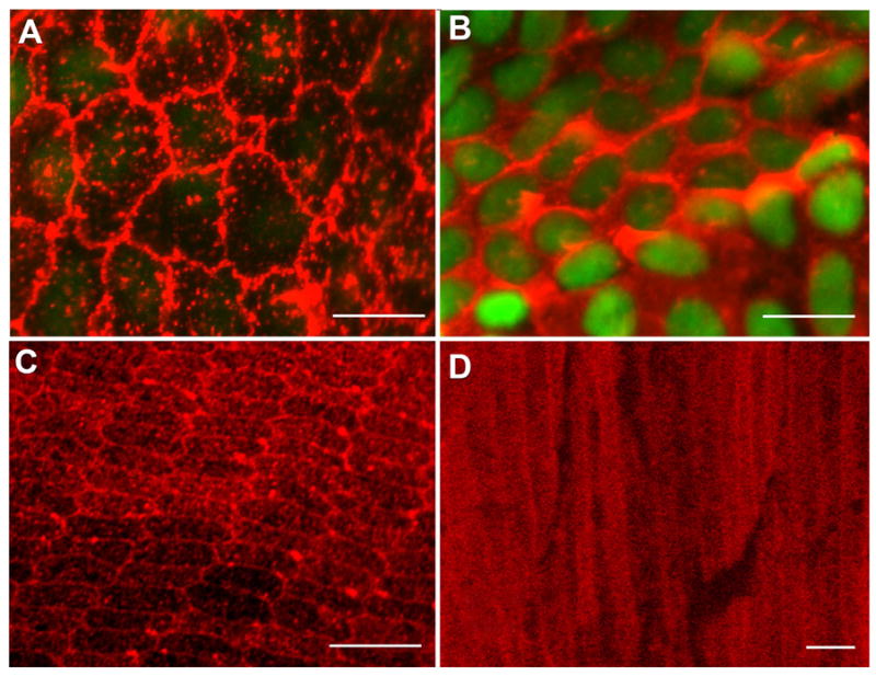
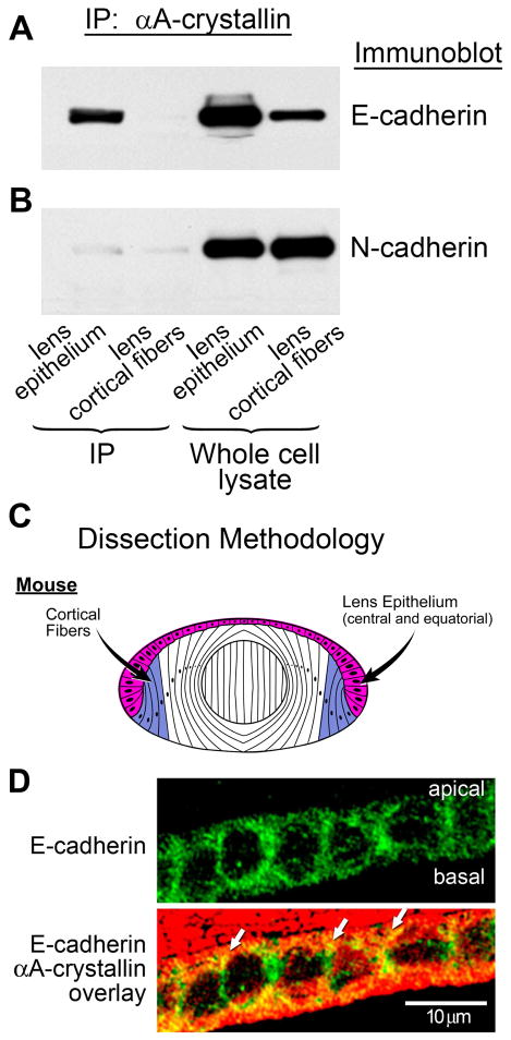
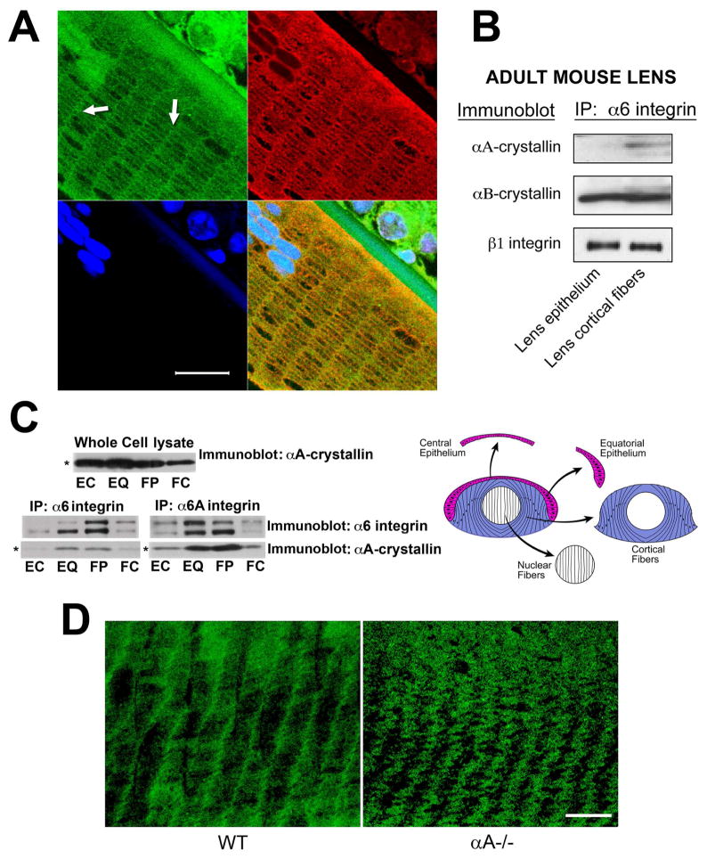
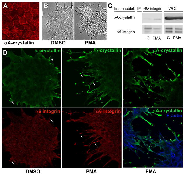
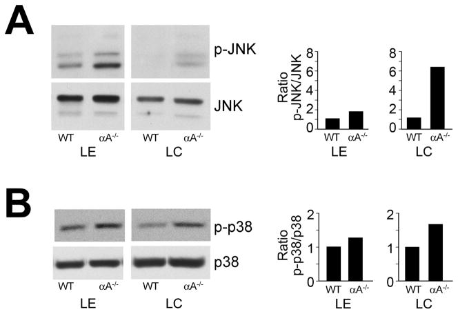
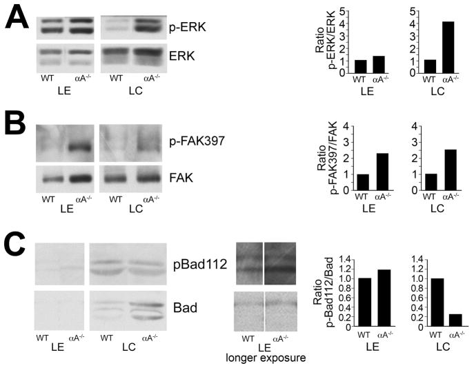
α-Crystallins are small heat-shock proteins important to lens transparency that provide the lens with its refractive properties. In their role as molecular chaperones, these crystallins also prevent protein aggregation, affect cytoskeletal remodeling, enhance resistance to cell stress, and provide lens cells with protection against apoptosis. While many of the functions assigned to αA-crystallin are attributable to its presence in the cytoplasm of lens cells, αA-crystallin also has been detected at the lens plasma membrane. However, how αA-crystallin becomes linked to the plasma membrane or what its functions are at this site has remained unknown. In this study, we examined the mechanisms by which αA-crystallin becomes associated with the lens membrane, focusing specifically on its interaction with membrane receptors, and the differentiation-specificity of these interactions. We also determined how the long-term absence of αA-crystallin alters receptor-linked signaling pathways. αA-crystallin association with membrane receptors was determined by co-immunoprecipitation analysis; its membrane localization was examined by confocal imaging; and the effect of αA-crystallin loss-of-function on the activation state of signaling molecules in pathways linked to membrane receptors was determined by immunoblot analysis. The results show that, in lens epithelial cells, plasma membrane αA-crystallin was primarily localized to apicolateral borders, reflecting the association of αA-crystallin with E-cadherin complexes. These studies also provide the first evidence that αA-crystallin maintained its association with the plasma membrane in lens cortical fiber cells, where it was localized to lateral interfaces, and further show that this association was mediated, in part, by αA-crystallin interaction with α6 integrin receptor complexes. We report that the absence of αA-crystallin led to constitutive activation of the stress kinases p38 and JNK, classical inducers of apoptotic cell death, and the loss of the phospho-Bad pro-survival signal, effects that were greatest in differentiating lens fiber cells. Concurrent with this, activation of FAK and ERK kinases was increased, demonstrating that these receptor-linked pathways also were dysregulated in the absence of αA-crystallin. These data link αA-crystallin plasma membrane association to its differentiation-state-specific interaction with E-cadherin and α6 integrin receptor complexes. The changes in cell signaling in αA-crystallin-null lenses suggest that dysregulation of receptor-linked cell-signaling pathways that accompany the failure of αA-crystallin to associate with membrane receptors may be responsible for the induction of apoptosis. The observed changes in lens cell signaling likely reflect long-term functional adaptations to the absence of the αA-crystallin chaperone/small heat-shock protein.


Figures

Figure 1
Figure 1
Confocal micrographs of αA-crystallin expression in the mouse lens. (A, B) Lens epithelial whole mounts prepared from adult wild-type mouse lenses were fixed and then incubated with a monoclonal antibody specific to αA-crystallin, followed by incubation with an Alexa568-conjugated secondary antibody (red); nuclei were stained with TOTO-1 (green). Cells were imaged by confocal microscopy at both apical (A) and basal (B) domains. Cells in the central epithelium are shown. (A) Note the expression of αA-crystallin on regions of plasma membrane at apical cell-cell interfaces. (B) Nuclei (green) are localized to the basal aspects of the lens epithelium where αA-crystallin was primarily cytoplasmic. Adult mouse lenses, ~150-day-old, were used to prepare lens epithelial whole mounts. (C) Cross-sectional and transverse sections of cortical lens fiber cells in adult wild-type mouse lenses. Cross-sectional slices were cut in the lens equatorial plane. Sections were stained with a monoclonal antibody to αA-crystallin and an Alexa568-conjugated secondary antibody (red). Intense staining for αA-crystallin was observed at the fiber cell interfaces in the outer cortical, hexagonally packed fiber cells. The periphery of the lens is at the bottom of the image. (D) In transverse sections the staining at cell-cell interfaces was shown to extend all along the lateral cell-cell borders of the cortical fiber cells. Cytoplasmic staining also was seen, which increased in the central fiber cells. Distance from the center of the lens is 400 μm. Scale bars = 10 μm (A), 20 μm (B, C), and 5μm (D).
Figure 2
Figure 2
Association of αA-crystallin with E-cadherin in lens epithelial cells. (A) Co-immunoprecipitation analysis of lens extracts following immunoprecipitation with an antibody to αA-crystallin, and immunoblotting with an antibody to E-cadherin. Note the strong association of αA-crystallin with E-cadherin in the lens epithelium, but no detectable association in lens cortical fibers. (B) Co-immunoprecipitation analysis of lens extracts following immunoprecipitation with an antibody to αA-crystallin and immunoblotting with an antibody to N-cadherin. Note that the association of N-cadherin with αA-crystallin was very low in both lens epithelium and lens cortical fiber cells. Whole cell lysates demonstrate the relative levels of E- and N-cadherin expression in lens epithelium and cortical fibers. (C) Diagram of dissection scheme for obtaining lens epithelial (red) and cortical fiber (blue) cells for these analyses from mouse lenses. In these preparations, the lens epithelial fraction comprised both central and equatorial epithelial cells. (D) Transverse sections of cortical lens fiber cells in adult wild-type mouse lenses were double stained for E-cadherin and αA-crystallin. E-cadherin was detected all along the lateral interfaces of these lens epithelial cells; αA-crystallin co-localized with E-cadherin (white arrows) particularly at the apical borders of these cells.
Figure 3
Figure 3
Differentiation-state-specific association of αA-crystallin with α6 integrin complexes. (A) Confocal microscopic analysis of cross-sections of cortical lens fiber cells double labeled with antibodies to α6 integrin and αA-crystallin. α6 integrin localized all along the cell-cell interfaces of these hexagonally-packed cortical fiber cells, but was predominantly associated with the short arms of the hexagon. αA-crystallin was co-localized with α6 integrin along these lateral interfaces (white arrows). The green staining of nuclei at the top left of the image is due to non-specific staining with the α6 integrin antibody. DNA was stained with draq5(blue). Scale bar = 20 μm. (B) Adult mouse lenses were dissected into epithelial and cortical fiber fractions as diagrammed in Figure 2C. Cell extracts were immunoprecipitated with an antibody to α6 integrin, and immunoblotted with antibodies to αA-crystallin, αB-crystallin, and ß1 integrin. Note that αA-crystallin association with α6 integrin was specific to cortical fiber cells, while the association of αB-crystallin with α6 integrin occurred in both lens epithelial and cortical fiber cells. (C) Chicken embryo lenses were dissected into four differentiation state-specific fractions: central epithelium (EC, red), equatorial epithelium (EQ, red), cortical fiber cells (FP, blue), and nuclear fiber cells (FC, white), as diagrammed in C. Cell extracts were immunoprecipitated either with an antibody to α6 integrin (recognizes both α6A and α6B integrin isoforms) or an antibody specific to α6A integrin, and immunoblotted with antibodies to αA-crystallin and α6 integrin. Note that the association of αA-crystallin with α6 integrin in these embryonic lenses was high in the zones of lens cell differentiation, including cells in the equatorial epithelium (EQ) and cortical fiber zones (FP). Note that the whole cell lysate (upper panel), had nearly equal levels of αA-crystallin in the central epithelium, equatorial epithelium and the cortical fiber cells. (D) Confocal microscopic analyses were performed on cortical fiber cells adjacent to the epithelium in wild-type (WT) and αA−/− lenses following immunostaining for α6 integrin. Note that α6 integrin was localized primarily to the short arms of these hexagonally packed fiber cells in wild-type lenses. While α6 integrin remained associated with the short arms of cortical fiber cells in αA−/− lenses, its linear organization at the plasma membrane was lost and these integrin junctions exhibited a wavy, disorganized pattern at the plasma membrane. Scale bar = 20 μm.
Figure 4
Figure 4
PMA activation of α6 integrin induces its association with αA-crystallin. (A) Confocal imaging of quail embryo lens epithelial cell cultures immunostained for αA-crystallin (red) showed αA-crystallin localized to cell-cell interfaces, demonstrating that these cells are a good model for studying the mechanism of αA-crystallin association with the plasma membrane. The dependence of αA-crystallin recruitment to α6 integrin on the activation state of this integrin was examined using PMA to activate α6 integrin. Control cultures were treated with DMSO. (B) Efficacy of PMA activation of the α6 integrin receptor was demonstrated by the elaboration of extensive lamellipodial/filopodial processes by the lens epithelial cells along a laminin coated substrate. (C) Recruitment of αA-crystallin to activated α6A integrin signaling complexes was determined following immunoprecipitation of α6 integrin with an antibody to the α6A subunit. Immunoprecipitates were immunoblotted with antibodies to both α6 integrin and αA-crystallin. PMA exposure induced αA-crystallin recruitment to α6 integrin complexes. (D) Co-localization of a-crystallins and α6 integrin in lamellipodial/filopodial processes extended in the presence of PMA was demonstrated by co-immunostaining. Cultures treated with either DMSO or PMA were double-labeled with antibodies to α6 integrin and α-crystallin (αA/αB-crystallin), or singly labeled with antibody to αA-crystallin and double stained with a nuclear marker. Both α-crystallins and α6 integrin were prominently localized to the processes the lens epithelial cells extended on the laminin substrate (arrows).
Figure 5
Figure 5
Activation of JNK and p38 stress pathways in αA−/− lens epithelial and fiber cells. Lens epithelial (LE) and cortical fiber cell (LC) fractions were dissected from 18–20 wild-type (WT) or αA−/− mouse lenses. Cell extracts were examined by immunoblot analysis with antibodies to (A) p-JNK and JNK, and (B) p-p38 and p38. Note that while p38 and JNK were activated in lens epithelial and cortical fiber cells of αA−/− mice, the degree of activation of these stress kinases was much greater in the cortical fiber zone of αA−/− lenses. Protein bands were quantified, and ratios of activated to total protein were determined and presented as fold-change between αA−/− and WT lenses. The results are representative of three independent experiments.
Figure 6
Figure 6
Changes in survival signaling in αA−/− lens epithelial and fiber cells. Lens epithelial (LE) and cortical fiber cell (LC) fractions were dissected from 18–20 wild-type or αA−/− mouse lenses. Cell extracts were examined by immunoblot analysis with antibodies to (A) p-ERK and ERK1/2, (B) p-FAK397 and FAK, and (C) pBad112 and Bad. The results show increased activation of both ERK and FAK in αA−/− lens epithelial and cortical fiber cells, but decreased phosphorylation of Bad in the cortical fiber zone. Protein bands were quantified, and the ratios of activated to total protein were determined and represented as fold-change between αA−/− and WT lenses. The results are representative of three independent experiments.
Figure 7
Figure 7
The αA-crystallin R116C mutant induces activation of p38 and FAK. Extracts of HLE B-3 lens epithelial cells transfected with either wild-type αA-crystallin or the αA-crystallin R116C mutant were examined by immunoblot analysis with antibodies to p-p38, p38, pFAK397, FAK, p-ERK, ERK, p-JNK, and β-actin. Expression of the αA-crystallin R116C mutant in lens epithelial cells induced significant activation of FAK and the stress kinase p38 compared with lens epithelial cells transfected with wild-type αA-crystallin. There was little difference in the activation state of JNK or ERK between αA-crystallin R116C- and WT-transfected lens epithelial cells.

Similar articles

Cited by

References

    1. Ahmed M, Forsberg J, Bergsten P. Protein profiling of human pancreatic islets by two-dimensional gel electrophoresis and mass spectrometry. J Proteome Res. 2005;4:931–940. - PubMed
    1. Andley UP. Crystallins in the eye: Function and pathology. Prog Retin Eye Res. 2007;26:78–98. - PubMed
    1. Andley UP, Mathur S, Griest TA, Petrash JM. Cloning, expression, and chaperone-like activity of human alphαA-crystallin. J Biol Chem. 1996;271:31973–31980. - PubMed
    1. Andley UP, Patel HC, Xi JH. The R116C mutation in alpha A-crystallin diminishes its protective ability against stress-induced lens epithelial cell apoptosis. J Biol Chem. 2002;277:10178–10186. - PubMed
    1. Andley UP, Rhim JS, Chylack LT, Jr, Fleming TP. Propagation and immortalization of human lens epithelial cells in culture. Invest Ophthalmol Vis Sci. 1994;35:3094–3102. - PubMed

Publication types

MeSH terms