

Visualizing the structural changes of bacteriophage Epsilon15 and its Salmonella host during infection
- PMID: 20709082
- PMCID: PMC3164490
- DOI: 10.1016/j.jmb.2010.07.058
Visualizing the structural changes of bacteriophage Epsilon15 and its Salmonella host during infection
Abstract
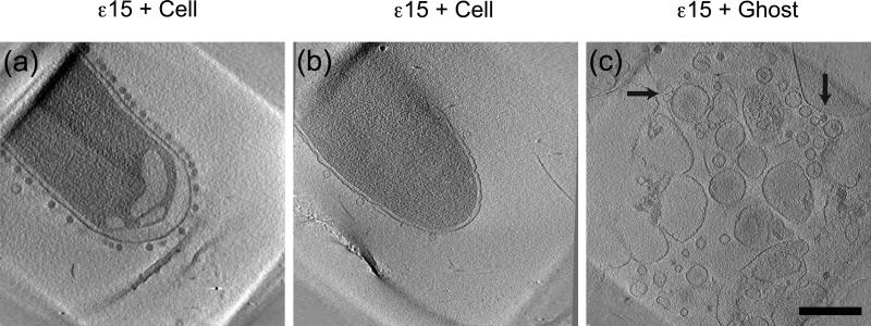
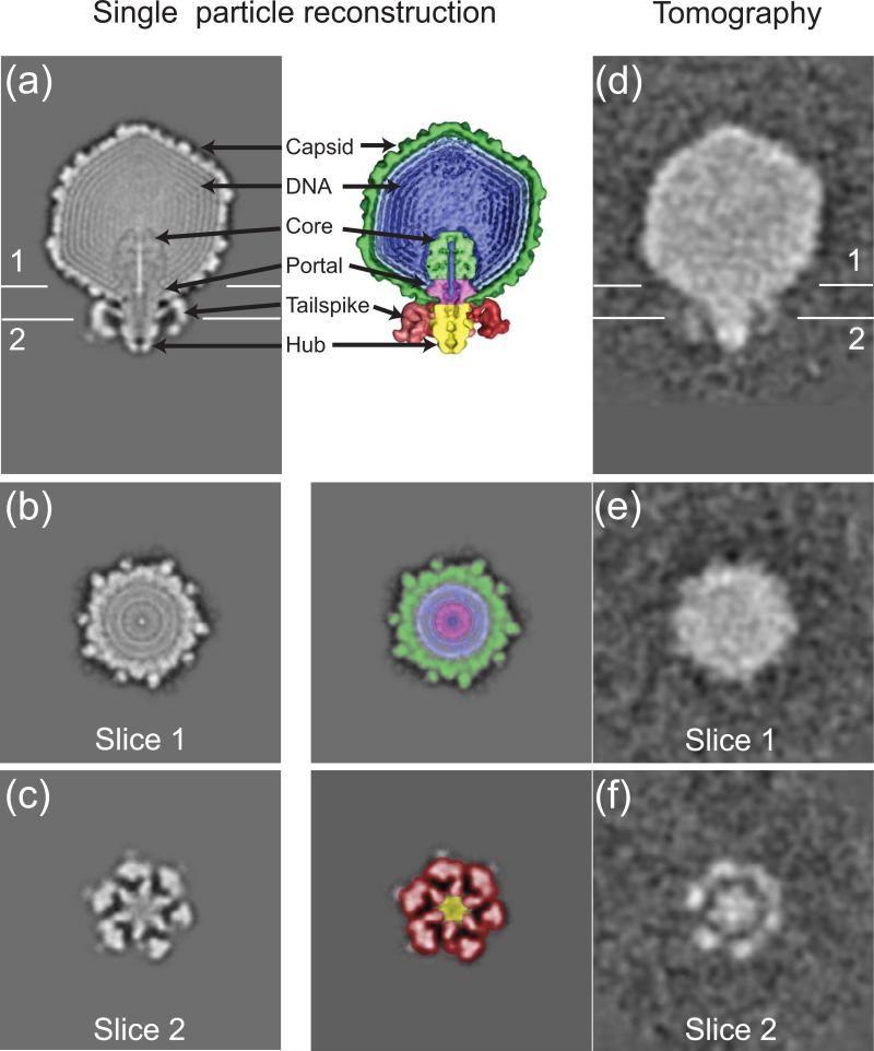
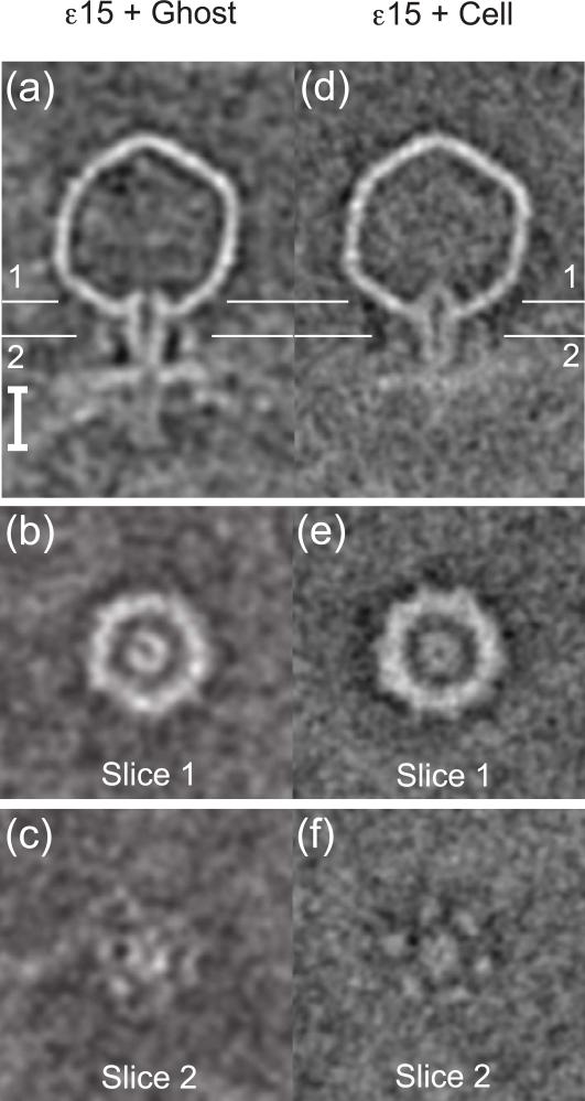
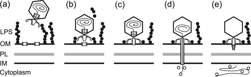
The efficient mechanism by which double-stranded DNA bacteriophages deliver their chromosome across the outer membrane, cell wall, and inner membrane of Gram-negative bacteria remains obscure. Advances in single-particle electron cryomicroscopy have recently revealed details of the organization of the DNA injection apparatus within the mature virion for various bacteriophages, including epsilon15 (ɛ15) and P-SSP7. We have used electron cryotomography and three-dimensional subvolume averaging to capture snapshots of ɛ15 infecting its host Salmonella anatum. These structures suggest the following stages of infection. In the first stage, the tailspikes of ɛ15 attach to the surface of the host cell. Next, ɛ15's tail hub attaches to a putative cell receptor and establishes a tunnel through which the injection core proteins behind the portal exit the virion. A tube spanning the periplasmic space is formed for viral DNA passage, presumably from the rearrangement of core proteins or from cellular components. This tube would direct the DNA into the cytoplasm and protect it from periplasmic nucleases. Once the DNA has been injected into the cell, the tube and portal seals, and the empty bacteriophage remains at the cell surface.
Copyright © 2010 Elsevier Ltd. All rights reserved.
Figures

References
-
- Simon LD, Anderson TF. The infection of Escherichia coli by T2 and T4 bacteriophages as seen in the electron microscope. I. Attachment and penetration. Virology. 1967;32:279–297. - PubMed
-
- Simon LD, Anderson TF. The infection of Escherichia coli by T2 and T4 bacteriophages as seen in the electron microscope. II. Structure and function of the baseplate. Virology. 1967;32:298–305. - PubMed
-
- Simon LD. The infection of Escherichia coli by T2 and T4 bacteriophages as seen in the electron microscope. 3. Membrane-associated intracellular bacteriophages. Virology. 1969;38:285–296. - PubMed
Publication types
MeSH terms
Substances
Grants and funding
LinkOut - more resources
Full Text Sources
