Calpain- and talin-dependent control of microvascular pericyte contractility and cellular stiffness
- PMID: 20709086
- PMCID: PMC2981705
- DOI: 10.1016/j.mvr.2010.07.012
Calpain- and talin-dependent control of microvascular pericyte contractility and cellular stiffness
Abstract
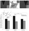
Pericytes surround capillary endothelial cells and exert contractile forces modulating microvascular tone and endothelial growth. We previously described pericyte contractile phenotype to be Rho GTPase- and α-smooth muscle actin (αSMA)-dependent. However, mechanisms mediating adhesion-dependent shape changes and contractile force transduction remain largely equivocal. We now report that the neutral cysteine protease, calpain, modulates pericyte contractility and cellular stiffness via talin, an integrin-binding and F-actin associating protein. Digital imaging and quantitative analyses of living cells reveal significant perturbations in contractile force transduction detected via deformation of silicone substrata, as well as perturbations of mechanical stiffness in cellular contractile subdomains quantified via atomic force microscope (AFM)-enabled nanoindentation. Pericytes overexpressing GFP-tagged talin show significantly enhanced contractility (~two-fold), which is mitigated when either the calpain-cleavage resistant mutant talin L432G or vinculin are expressed. Moreover, the cell-penetrating, calpain-specific inhibitor termed CALPASTAT reverses talin-enhanced, but not Rho GTP-dependent, contractility. Interestingly, our analysis revealed that CALPASTAT, but not its inactive mutant, alters contractile cell-driven substrata deformations while increasing mechanical stiffness of subcellular contractile regions of these pericytes. Altogether, our results reveal that calpain-dependent cleavage of talin modulates cell contractile dynamics, which in pericytes may prove instrumental in controlling normal capillary function or microvascular pathophysiology.
Copyright © 2010 Elsevier Inc. All rights reserved.
Figures


Similar articles
-
Pericyte Rho GTPase mediates both pericyte contractile phenotype and capillary endothelial growth state.Am J Pathol. 2007 Aug;171(2):693-701. doi: 10.2353/ajpath.2007.070102. Epub 2007 Jun 7. Am J Pathol. 2007. PMID: 17556591 Free PMC article.
-
Pericyte actomyosin-mediated contraction at the cell-material interface can modulate the microvascular niche.J Phys Condens Matter. 2010 May 19;22(19):194115. doi: 10.1088/0953-8984/22/19/194115. Epub 2010 Apr 26. J Phys Condens Matter. 2010. PMID: 21386441
-
Talin contains a C-terminal calpain2 cleavage site important in focal adhesion dynamics.PLoS One. 2012;7(4):e34461. doi: 10.1371/journal.pone.0034461. Epub 2012 Apr 4. PLoS One. 2012. PMID: 22496808 Free PMC article.
-
Pericyte-Endothelial Interactions in the Retinal Microvasculature.Int J Mol Sci. 2020 Oct 8;21(19):7413. doi: 10.3390/ijms21197413. Int J Mol Sci. 2020. PMID: 33049983 Free PMC article. Review.
-
The pericyte: cellular regulator of microvascular blood flow.Microvasc Res. 2009 May;77(3):235-46. doi: 10.1016/j.mvr.2009.01.007. Epub 2009 Feb 7. Microvasc Res. 2009. PMID: 19323975 Free PMC article. Review.
Cited by
-
Calpain inhibition preserves talin and attenuates right heart failure in acute pulmonary hypertension.Am J Respir Cell Mol Biol. 2012 Sep;47(3):379-86. doi: 10.1165/rcmb.2011-0286OC. Epub 2012 May 10. Am J Respir Cell Mol Biol. 2012. PMID: 22582173 Free PMC article.
-
The Adhesome Network: Key Components Shaping the Tumour Stroma.Cancers (Basel). 2021 Jan 30;13(3):525. doi: 10.3390/cancers13030525. Cancers (Basel). 2021. PMID: 33573141 Free PMC article. Review.
-
Pericyte-endothelial crosstalk: implications and opportunities for advanced cellular therapies.Transl Res. 2014 Apr;163(4):296-306. doi: 10.1016/j.trsl.2014.01.011. Epub 2014 Jan 24. Transl Res. 2014. PMID: 24530608 Free PMC article. Review.
-
Attenuation of Blood-Brain Barrier Breakdown and Hyperpermeability by Calpain Inhibition.J Biol Chem. 2016 Dec 30;291(53):26958-26969. doi: 10.1074/jbc.M116.735365. Epub 2016 Nov 8. J Biol Chem. 2016. PMID: 27875293 Free PMC article.
-
Pericytes derived from adipose-derived stem cells protect against retinal vasculopathy.PLoS One. 2013 May 31;8(5):e65691. doi: 10.1371/journal.pone.0065691. Print 2013. PLoS One. 2013. PMID: 23741506 Free PMC article.
References
-
- Aebi U, Pollard TD. A glow discharge unit to render electron microscope grids and other surfaces hydrophilic. J. Electron Microsc. Tech. 1987;7:29–33. - PubMed
-
- Balaban NQ, Schwarz US, Riveline D, Goichberg P, Tzur G, Sabanay I, Mahalu D, Safran S, Bershadsky A, Addadi L, Geiger B. Force and focal adhesion assembly: a close relationship studied using elastic micropatterned substrates. Nat. Cell Biol. 2001;3:466–472. - PubMed
Publication types
MeSH terms
Substances
Grants and funding
LinkOut - more resources
Full Text Sources
Miscellaneous

