Comparative Study
. 2010 Dec;97(2):227-38.
doi: 10.1016/j.pbb.2010.08.003. Epub 2010 Aug 13.

The effect of restraint stress on prepulse inhibition and on corticotropin-releasing factor (CRF) and CRF receptor gene expression in Wistar-Kyoto and Brown Norway rats

Affiliations
Comparative Study

The effect of restraint stress on prepulse inhibition and on corticotropin-releasing factor (CRF) and CRF receptor gene expression in Wistar-Kyoto and Brown Norway rats

Jane E Sutherland et al. Pharmacol Biochem Behav. 2010 Dec.

Abstract

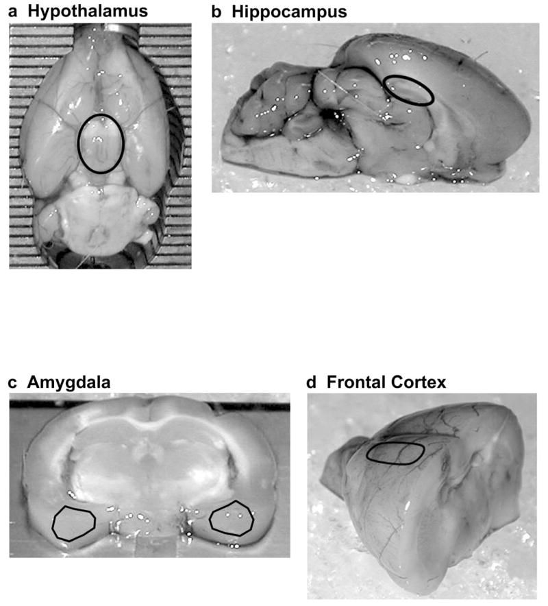
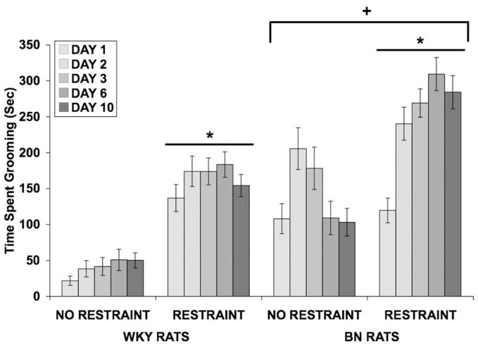
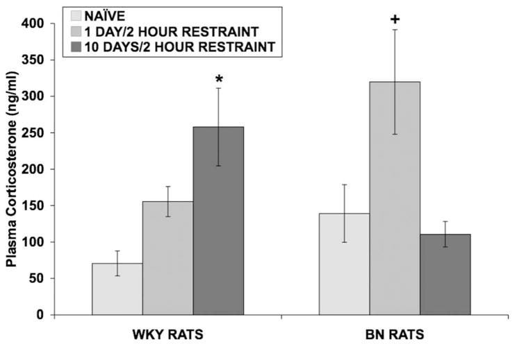
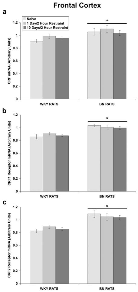
Stress plays a role in many psychiatric disorders that are characterized by deficits in prepulse inhibition (PPI), a form of sensorimotor gating. Corticotropin-releasing factor (CRF) is one of the most important neurotransmitters involved in behavioral components of the stress response, and central infusion of CRF decreases PPI in rodents. We recently demonstrated that restraint stress decreases PPI and attenuates the increase in PPI caused by repeated testing. To broaden our investigation into how restraint affects PPI, we subjected Wistar-Kyoto (WKY) and Brown Norway (BN) rats to 10 consecutive days of 2-hour restraint, or to brief handling, prior to assessing PPI. We next examined the effects of 1 or 10days of 2-hour restraint on plasma corticosterone levels in order to determine whether the endocrine response to stress parallels the behavioral effect of stress. Finally, we examined the effects of 1 or 10days of 2-hour restraint on CRF and CRF receptor gene expression in the amygdala, hippocampus, frontal cortex, and hypothalamus in order to determine whether a temporal pattern of gene expression parallels the change in the behavioral response to stress. The major findings of the present study are that 1) restraint stress attenuates the increase in PPI caused by repeated testing in both WKY and BN rats, and BN rats are more sensitive to the effects of restraint on PPI than WKY rats, 2) restraint-induced increases in corticosterone levels mirror the effect of restraint on PPI in WKY rats but not in BN rats, 3) laterality effects on gene expression were observed for the amygdala, whereby restraint increases CRF gene expression in the left, but not right, amygdala, and 4) some restraint-induced changes in CRF and CRF receptor gene expression precede changes in PPI while other changes coincide with altered PPI in a rat strain- and brain region-dependent manner.


Figures

Fig 1
Fig 1
Regions of the brain used for assessing mRNA levels. (a) The hypothalamus (black circle) was extracted from the ventral surface of the rat brain. (b) The hippocampi (black circle) were dissected from a mid-sagittal section of the rat brain. (c) 2.0 mm thick sections of amygdalae (black triangles) were dissected from a coronal section of the rat brain. (d) Frontal cortices (black oval) were dissected from an anterior portion of the rat brain. Dissection techniques and approximate stereotaxic coordinates for these brain regions are described in greater detail under “Brain Dissection” in the Methods.
Fig 2
Fig 2
Effect of restraint stress on PPI in WKY and BN rats. Values shown are means ± SEMs. For all groups, n = 15–16. Rats were restrained for 2 hours, once a day for 10 consecutive days, or were handled briefly and returned to the home cage. PPI was assessed 30 minutes after restraint termination on days 1, 2, 3, 6, and 10. Prepulse stimulus intensities are 76, 82, 85, and 88 dB. (a) On day 1, restraint did not alter PPI in WKY or BN rats. (b) On day 2, restraint did not alter PPI in WKY or BN rats. (c) Exposure to 3 days of 2-hour restraint significantly attenuated the increase in PPI caused by repeated testing in BN rats (*p = 0.016). (d) Exposure to 6 days of 2-hour restraint also significantly attenuated the increase in PPI caused by repeated testing in BN rats (*p = 0.003). (e) Exposure to 10 days of 2-hour restraint significantly attenuated the increase in PPI caused by repeated testing in WKY (+p = 0.030) and BN rats (*p = 0.037).
Fig 3
Fig 3
Effect of restraint stress on baseline startle amplitude in WKY and BN rats. Values shown are means ± SEMs and were calculated from the startle stimulus alone trials that were used to calculate percent PPI. Startle amplitude was assessed 30 minutes after restraint termination on days 1, 2, 3, 6, and 10. BN rats exhibited a lower startle response (*p < 0.001 vs. WKY rats). Restraint did not alter startle amplitude in either strain, on any day examined.
Fig 4
Fig 4
Effect of restraint stress on time spent grooming (in seconds) in WKY and BN rats. Values shown are means ± SEMs. For all groups, n = 15–16. Grooming was assessed for 15 minutes, beginning immediately after restraint termination on days 1, 2, 3, 6, and 10. Restraint increased time spent grooming in both rat strains (*p < 0.001 vs. No Restraint). BN rats spent more time grooming overall (+p < 0.001 vs. WKY rats).
Fig 5
Fig 5
Effect of restraint stress on plasma corticosterone levels in WKY and BN rats. Values shown are means ± SEMs. For all groups, n = 5. Rats were exposed to either: 1) brief handling (naïve controls), 2) one day of 2-hour restraint, or 3) ten consecutive days of 2-hour restraint. Rats were rapidly decapitated and trunk blood was collected 30 minutes after termination of the last session of restraint. In WKY rats, 10 days of 2-hour restraint increased corticosterone levels (*p = 0.006 vs. Naïve WKY rats, Tukey’s test). In BN rats, 1 day of 2-hour restraint increased corticosterone levels (+p 0.053 vs. Naïve BN rats, Tukey’s test).
Fig 6
Fig 6
Effect of restraint stress on CRF and CRF receptor mRNA levels in the left and right amygdala of WKY and BN rats. Values are expressed as a ratio of CRF mRNA to β-glucuronidase mRNA and CRF1 (or CRF2) receptor mRNA to β-2 microglobulin mRNA in arbitrary units (means ± SEMs). For all groups, n = 5–7. Rats were exposed to either: 1) brief handling (naïve controls), 2) one day of 2-hour restraint, or 3) ten consecutive days of 2-hour restraint. Thirty minutes after termination of the last session of restraint, rats were rapidly decapitated and the amygdala was dissected. Note: the scale on the y-axis differs between CRF1 and CRF2 receptor mRNA graphs. (a) 1 day of 2-hour restraint increased CRF mRNA levels in the left amygdala of WKY and BN rats (*p = 0.012 vs. Naïve rats, Tukey’s test). In BN rats, CRF mRNA levels returned to baseline after 10 days of 2-hour restraint (+p = 0.027 vs. 1 Day/2 Hour Restraint, Tukey’s test). Restraint did not alter CRF mRNA levels in the right amygdala of either rat strain. (b) In the right hemisphere, BN rats had greater levels of CRF1 receptor mRNA (*p = 0.010 vs. WKY rats). (c) Restraint stress did not significantly alter CRF2 receptor mRNA levels in either the left or right amygdala of either rat strain.
Fig 7
Fig 7
Effect of restraint stress on CRF and CRF receptor mRNA levels in the hippocampus of WKY and BN rats. Values are expressed as a ratio of CRF mRNA to β-glucuronidase mRNA and CRF1 (or CRF2) receptor mRNA to β-2 microglobulin mRNA in arbitrary units (means ± SEMs). For all groups, n = 4–5. Rats were exposed to either: 1) brief handling (naïve controls), 2) one day of 2-hour restraint, or 3) ten consecutive days of 2-hour restraint. Thirty minutes after termination of the last session of restraint, rats were rapidly decapitated and the hippocampus was dissected. Note: the scale on the y-axis differs between CRF1 and CRF2 receptor mRNA graphs. (a) In BN rats, both 1 day (*p < 0.001 vs. Naïve BN rats, Tukey’s test) and 10 days (+p = 0.008 vs. Naïve BN rats, Tukey’s test) of restraint increased CRF mRNA levels. (b) In BN rats, 10 days of restraint increased CRF1 receptor mRNA levels (*p = 0.007 vs. Naïve BN rats, Tukey’s test). (c) Restraint did not affect CRF2 receptor mRNA levels in either rat strain.
Fig 8
Fig 8
Effect of restraint stress on CRF and CRF receptor mRNA levels in the frontal cortex of WKY and BN rats. Values are expressed as a ratio of CRF mRNA to β-glucuronidase mRNA and CRF1 (or CRF2) receptor mRNA to β-2 microglobulin mRNA in arbitrary units (means ± SEMs). For all groups, n = 5–7. Rats were exposed to either: 1) brief handling (naïve controls), 2) one day of 2-hour restraint, or 3) ten consecutive days of 2-hour restraint. Thirty minutes after termination of the last session of restraint, rats were rapidly decapitated and the frontal cortex was dissected. (a) BN rats had greater levels of CRF mRNA in the frontal cortex (*p = 0.004 vs. WKY rats). (b) BN rats had greater levels of CRF1 receptor mRNA (*p < 0.001 vs. WKY rats). (c) BN rats had greater levels of CRF2 receptor mRNA (*p < 0.001 vs. WKY rats).
Fig 9
Fig 9
Effect of restraint stress on CRF and CRF receptor mRNA levels in the hypothalamus of WKY and BN rats. Values are expressed as a ratio of CRF mRNA to β-glucuronidase mRNA and CRF1 (or CRF2) receptor mRNA to β-2 microglobulin mRNA in arbitrary units (means ± SEMs). For all groups, n = 5. Rats were exposed to either: 1) brief handling (naïve controls), 2) one day of 2-hour restraint, or 3) ten consecutive days of 2-hour restraint. Thirty minutes after termination of the last session of restraint, rats were rapidly decapitated and the hypothalamus was dissected. Note: the scale on the y-axis differs between CRF1 and CRF2 receptor mRNA graphs. (a) 10 days of 2-hour restraint increased CRF mRNA levels in BN rats (*p = 0.022 vs. Naïve BN rats, Tukey’s test). (b) Naïve BN rats had greater levels of CRF1 receptor mRNA (*p = 0.050 vs. Naïve WKY rats, independent t-test). (c) Naïve BN rats showed a trend towards having greater levels of CRF2 receptor mRNA (*p = 0.053 vs. Naïve WKY rats, independent t-test).

Similar articles

Cited by

References

    1. Alsene KM, Carasso BS, Connors EE, Bakshi VP. Disruption of prepulse inhibition after stimulation of central but not peripheral alpha-1 adrenergic receptors. Neuropsychopharmacology. 2006;31:2150–61. - PubMed
    1. Baker KB, Kim JJ. Amygdalar lateralization in fear conditioning: evidence for greater involvement of the right amygdala. Behav Neurosci. 2004;118:15–23. - PubMed
    1. Balagura S, Harrell LE, Roy E. Effect of the light-dark cycle on neuroendocrine and behavioral responses to scheduled feeding. Physiol Behav. 1975;15:245–7. - PubMed
    1. Bale TL, Vale WW. CRF and CRF receptors: role in stress responsivity and other behaviors. Annu Rev Pharmacol Toxicol. 2004;44:525–57. - PubMed
    1. Braff D, Stone C, Callaway E, Geyer M, Glick I, Bali L. Prestimulus effects on human startle reflex in normals and schizophrenics. Psychophysiology. 1978;15:339–43. - PubMed

Publication types

MeSH terms

Substances