. 2010 Nov;169(2):365-74.
doi: 10.1016/j.jviromet.2010.08.006. Epub 2010 Aug 13.

Generation of VSV pseudotypes using recombinant ΔG-VSV for studies on virus entry, identification of entry inhibitors, and immune responses to vaccines

Affiliations

Generation of VSV pseudotypes using recombinant ΔG-VSV for studies on virus entry, identification of entry inhibitors, and immune responses to vaccines

Michael A Whitt. J Virol Methods. 2010 Nov.

Abstract

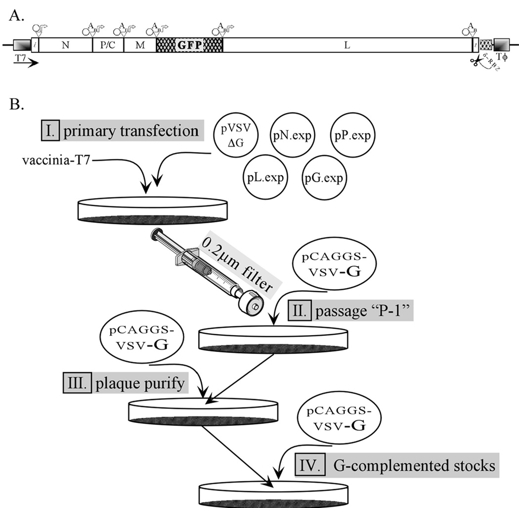
Vesicular stomatitis virus (VSV) is a prototypic enveloped animal virus that has been used extensively to study virus entry, replication and assembly due to its broad host range and robust replication properties in a wide variety of mammalian and insect cells. Studies on VSV assembly led to the creation of a recombinant VSV in which the glycoprotein (G) gene was deleted. This recombinant (rVSV-ΔG) has been used to produce VSV pseudotypes containing the envelope glycoproteins of heterologous viruses, including viruses that require high-level biocontainment; however, because the infectivity of rVSV-ΔG pseudotypes is restricted to a single round of replication the analysis can be performed using biosafety level 2 (BSL-2) containment. As such, rVSV-ΔG pseudotypes have facilitated the analysis of virus entry for numerous viral pathogens without the need for specialized containment facilities. The pseudotypes also provide a robust platform to screen libraries for entry inhibitors and to evaluate the neutralizing antibody responses following vaccination. This manuscript describes methods to produce and titer rVSV-ΔG pseudotypes. Procedures to generate rVSV-ΔG stocks and to quantify virus infectivity are also described. These protocols should allow any laboratory knowledgeable in general virological and cell culture techniques to produce successfully replication-restricted rVSV-ΔG pseudotypes for subsequent analysis.


Figures

Figure 1
Figure 1
Recovery, growth and analysis of rVSV-ΔG. (A) Diagram of the cDNA encoding the anti-genome of rVSV-ΔG-GFP. The conserved VSV transcriptional regulatory sequences (octagon = stop sequence; An= polyadenylation sequence; arrow = transcriptional promoter or start sequence) are shown above the diagram. The bacteriophage T7 RNA polymerase promoter (T7) and terminator sequences (Tϕ) and the hepatitis delta virus ribozyme (δ-RBZ) with cleavage site are indicated below the diagram. (B) A flow chart illustrating the key points involved in generating and characterizing recombinant rVSV-ΔG. (I) Initially cells are cotransfected with the plasmid pVSV-ΔG-GFP and the four support plasmids encoding the VSV N, P, G and L proteins. This is followed by virus propagation (II), with subsequent plaque-purification and growth of G-complemented rVSV-ΔG-GFP working stocks (III & IV). The G-complemented virus stocks produced in step IV are titrated, and then used to infect cells that express a heterologous glycoprotein for production of the ΔG-GFP pseudotypes. Figure courtesy of C.S. Robison (Robison, 2001).
Figure 2
Figure 2
Phase contrast micrograph of BHK-21 cells taken immediately before starting a recovery experiment for rVSV-ΔG viruses. The cells should be evenly distributed and should ~90–95% confluent. The image was collected using a digital camera connected to a phototube with a 2.5X lens on an Olympus CK2 microscope equipped with a 10X phase objective. The image brightness was adjusted in Canvas 11 using the “levels” function to optimize visualization of individual cells.
Figure 3
Figure 3
Phase contrast images of two fields of cells at 22 hours post-transfection with pCAGGS-G. Arrowheads indicate regions of small syncytia beginning to form. Images were captured and adjusted to optimize cell visualization as described for Fig. 2.
Figure 4
Figure 4
Immunofluorescence/phase contrast micrograph of ΔL-GFP transfected cells 24 hours post-transfection. Cells were imaged using low-level brightfield illumination and fluorescence was achieved by excitation using a FITC-filter set on a Zeiss Axiophot using Axiovision software. Quantification of GFP-positive cells revealed that 21.5% of the cells were replicating ΔL-GFP.
Figure 5
Figure 5
Phase contrast images of cells 24 or 48 hours post-transfer of two independent supernatants from a recovery experiment. The cells had been transfected with pCAGGS-G 24 hours prior to transfer of 0.2 µ filtered recovery supernatants. The top panels show cells after transfer of the supernatant from a culture that was infected with vvT7 and transfected with only the support plasmids. The middle and lower panels are from two independent recovery experiments. The middle panel has a small area of infected cells (arrow) near the middle of the image which are showing VSV-induced CPE. The lower panel also has a small, but much less obvious area of CPE (arrowhead). By 48 hours cells that were infected with the two recovery trial supernatants show extensive cell rounding, characteristic of VSV-induced CPE while the mock culture (top, right panel) does not.

Similar articles

Cited by

References

    1. Abe K, Nozaki A, Tamura K, Ikeda M, Naka K, Dansako H, Hoshino HO, Tanaka K, Kato N. Tandem repeats of lactoferrin-derived anti-hepatitis C virus peptide enhance antiviral activity in cultured human hepatocytes. Microbiol. Immunol. 2007;51:117–125. - PubMed
    1. Fredericksen BL, Whitt MA. Vesicular stomatitis virus glycoprotein mutations that affect membrane fusion activity and abolish virus infectivity. J. Virol. 1995;69:1435–1443. - PMC - PubMed
    1. Fuerst TR, Niles EG, Studier RW, Moss B. Eukaryotic transient-expression system based on recombinant vaccinia virus that synthesizes bacteriophage T7 RNA polymerase. Proc.Natl.Acad.Sci.USA. 1986;83:8122–8126. - PMC - PubMed
    1. Fukushi S, Mizutani T, Saijo M, Matsuyama S, Miyajima N, Taguchi F, Itamura S, Kurane I, Morikawa S. Vesicular stomatitis virus pseudotyped with severe acute respiratory syndrome coronavirus spike protein. J. Gen. Virol. 2005;86:2269–2274. - PubMed
    1. Fukushi S, Watanabe R, Taguchi F. Pseudotyped vesicular stomatitis virus for analysis of virus entry mediated by SARS coronavirus spike proteins. Methods Mol. Biol. 2008;454:331–338. - PMC - PubMed

Publication types