. 2010 Nov 10;170(4):1045-55.
doi: 10.1016/j.neuroscience.2010.08.012. Epub 2010 Aug 13.

Membrane estrogen receptors activate the metabotropic glutamate receptors mGluR5 and mGluR3 to bidirectionally regulate CREB phosphorylation in female rat striatal neurons

Affiliations

Membrane estrogen receptors activate the metabotropic glutamate receptors mGluR5 and mGluR3 to bidirectionally regulate CREB phosphorylation in female rat striatal neurons

D Grove-Strawser et al. Neuroscience. .

Abstract

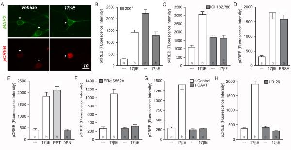
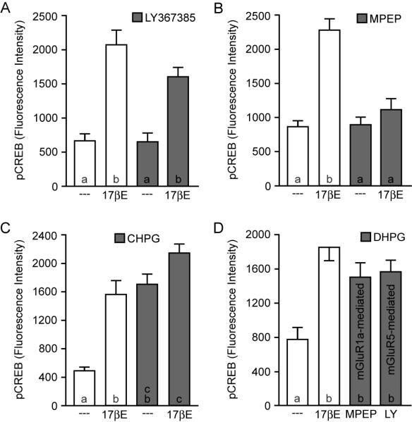
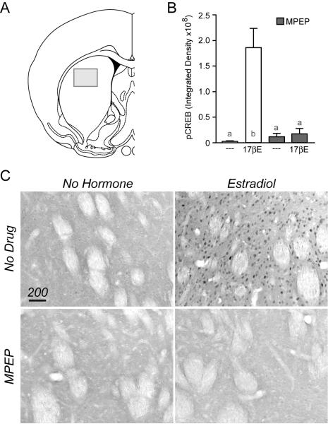
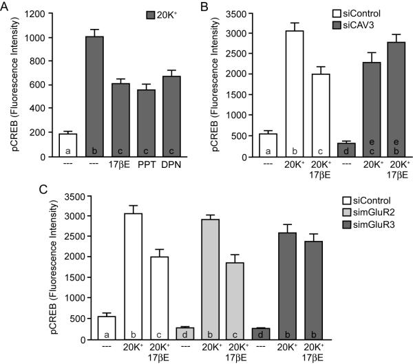
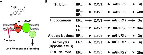
Along with its ability to directly regulate gene expression, estradiol influences cell signaling and brain functions via rapid, membrane-initiated events. In the female rat striatum, estradiol activates membrane-localized estrogen receptors to influence synaptic neurotransmission, calcium channel activity, and behaviors related to motor control. Yet, the mechanism by which estradiol acts to rapidly affect striatal physiology has remained elusive. Here we find that membrane estrogen receptors (ERs) couple to the metabotropic glutamate receptors mGluR5 and mGluR3, providing the framework to understand how membrane estrogen receptors affect striatal function. Using CREB phosphorylation as a downstream measure of ER/mGluR activation, membrane-localized estrogen receptor α (ERα) activates mGluR5 signaling to mediate mitogen-activated protein kinase (MAPK)-dependent CREB phosphorylation. Further, ERα and estrogen receptor β (ERβ) activate mGluR3 to attenuate L-type calcium channel-dependent CREB signaling. Interestingly, while this fundamental mechanism of ER/mGluR signaling was initially characterized in hippocampal neurons, estrogen receptors in striatal neurons are paired with a different set of mGluRs, resulting in the potential to functionally isolate membrane-initiated estrogen signaling across brain regions via use of specific mGluR modulators. These results provide both a mechanism for the rapid actions of estrogens within the female striatum, as well as demonstrate that estrogen receptors can interact with a more diverse set of surface membrane receptors than previously recognized.


Figures

Figure 1
Figure 1
Estradiol-induced CREB phosphorylation in female striatal neurons. (A) Confocal images of cultured neurons immunolabeled with Microtubule Associated Protein 2 (MAP2; green) and CREB, phosphorylated at serine 133 (pCREB; red). In comparison to vehicle controls, striatal neurons treated with estradiol (17βE; 1 nM) for five minutes exhibit increased staining for pCREB (scale bar, 10 μm). Arrowheads indicate nuclei of neurons. (B) Quantification of pCREB immunolabeling following estradiol and/or 20 mM K+ stimulation (F = 38.65; letters within the bars indicate statistically different groups). While estradiol alone increased CREB phosphorylation (open bars), estradiol attenuated the rise in CREB phosphorylation mediated by a three-minute depolarization with 20 mM K+ (filled bars). (C) Pretreatment of striatal cultures with the estrogen receptor antagonist, ICI 182,780 (ICI; 100 nM), eliminated estradiol-induced CREB phosphorylation (F = 31.26). (D) The membrane-impermeable estrogen analog, estradiol conjugated to bovine serum albumen (EBSA) mimicked the actions of estradiol (F = 29.46). (E) Activation of ERα with the receptor agonist PPT (1 nM) mimicked the actions of estradiol, whereas the ERβ-specific agonist DPN (10 nM) was without effect (F = 60.08). (F-H) The actions of estradiol on CREB phosphorylation could be blocked by inhibiting the trafficking of ERα to the surface membrane via overexpression of EGFP-S522A (F; F = 41.28), disruption of caveolin-1 (CAV1) expression by means of siRNA knockdown (G; F = 41.28) or inhibition of MAPK signaling by pretreating the cultures with the MEK inhibitor U0126 (10 μM) (H; F = 159.9).
Figure 2
Figure 2
Estradiol-mediated CREB phosphorylation occurs via selective activation of mGluR5. (A-B) In contrast to cells from hippocampus and other brain regions, estradiol-induced CREB phosphorylation in striatal neurons is not blocked by the mGluR1a antagonist LY367385 (100 μM) (F = 21.56). Rather, estradiol-mediated CREB phosphorylation in striatal neurons is eliminated by the mGluR5 antagonist MPEP (5 μM) (F = 24.67). (C) The mGluR5 agonist CHPG (1 mM) increased CREB phosphorylation similar to estradiol, and occluded the effect of the steroid (F = 24.93). (D) Application of the pan-specific group I mGluR agonist DHPG (50 μM) in the presence of either a mGluR5 (MPEP) or a mGluR1a (LY367385) specific antagonist resulted in an increase in CREB phosphorylation, suggesting that while ERα activates mGluR5 in striatal neurons, both group I mGluRs are functionally linked to CREB signaling in this system.
Figure 3
Figure 3
Estradiol induces CREB phosphorylation via mGluR5 in rat striatum. (A) Illustration depicting the approximate location within the striatum in which pCREB staining was quantified. (B) CREB phosphorylation was significantly increased 15 minutes following an injection (s.c.) of estradiol when compared to vehicle controls. This effect of estradiol was blocked by the mGluR5 antagonist MPEP, injected (i.p.) 20 min prior to hormone exposure (F = 9.37). (C) Example immunohistochemical images of pCREB in striatal tissue.
Figure 4
Figure 4
Estradiol attenuation of L-type calcium channel-dependent CREB phosphorylation in striatal neurons is dependent on mGluR3. (A) Activation of either ERα or ERβ with PPT or DPN mimicked the actions of estradiol on attenuating L-type calcium channel-dependent CREB phosphorylation (F = 43.69). (B) Knockdown of caveolin-3 (CAV3) expression eliminated estradiol regulation of L-type calcium channel signaling (F = 49.86). (C) In striatal neurons, disruption of mGluR3, but not mGluR2 expression blocked the actions of estradiol on L-type calcium channel-mediated CREB phosphorylation (F = 54.93).
Figure 5
Figure 5
Brain region specificity in estradiol-induced activation of mGluRs. (A) General depiction of the proposed mechanism by which caveolin proteins act to functionally isolate distinct estrogen receptors and mGluRs, leading to activation of specific second messenger signaling cascades. (B) In striatal neurons, ERα is functionally coupled to mGluR5 via CAV1. In addition, a separate pool of ERα and ERβ are coupled to mGluR3 via expression of CAV3. This is in contrast to neurons of the hippocampus, arcuate nucleus and DRG, as well as astrocytes of the hypothalamus in which estrogen receptors appear principally coupled to mGluR1a and/or mGluR2. Grayed text indicates preparations in which the caveolin protein responsible for ER coupling to mGluRs was not determined.

Similar articles

Cited by

References

    1. Augustine GJ, Santamaria F, Tanaka K. Local calcium signaling in neurons. Neuron. 2003;40:331–346. - PubMed
    1. Balthazart J, Ball GF. Is brain estradiol a hormone or a neurotransmitter? Trends Neurosci. 2006;29:241–249. - PubMed
    1. Becker JB. Direct effect of 17 beta-estradiol on striatum: sex differences in dopamine release. Synapse. 1990;5:157–164. - PubMed
    1. Becker JB, Snyder PJ, Miller MM, Westgate SA, Jenuwine MJ. The influence of estrous cycle and intrastriatal estradiol on sensorimotor performance in the female rat. Pharmacol Biochem Behav. 1987;27:53–59. - PubMed
    1. Bondar G, Kuo J, Hamid N, Micevych P. Estradiol-induced estrogen receptor-alpha trafficking. J Neurosci. 2009;29:15323–15330. - PMC - PubMed

Publication types

MeSH terms

Substances