. 2010 Aug 19;8(2):186-95.
doi: 10.1016/j.chom.2010.07.007.

Direct complement restriction of flavivirus infection requires glycan recognition by mannose-binding lectin

Affiliations

Direct complement restriction of flavivirus infection requires glycan recognition by mannose-binding lectin

Anja Fuchs et al. Cell Host Microbe. .

Abstract

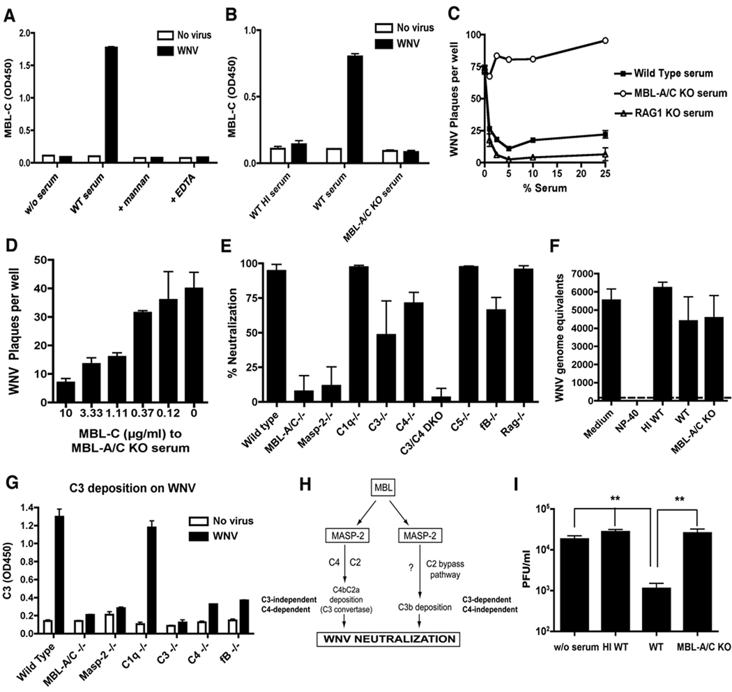
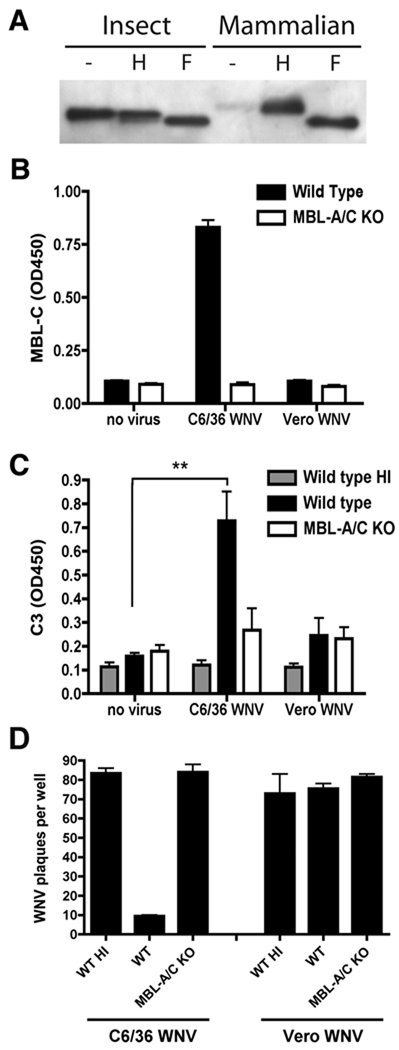
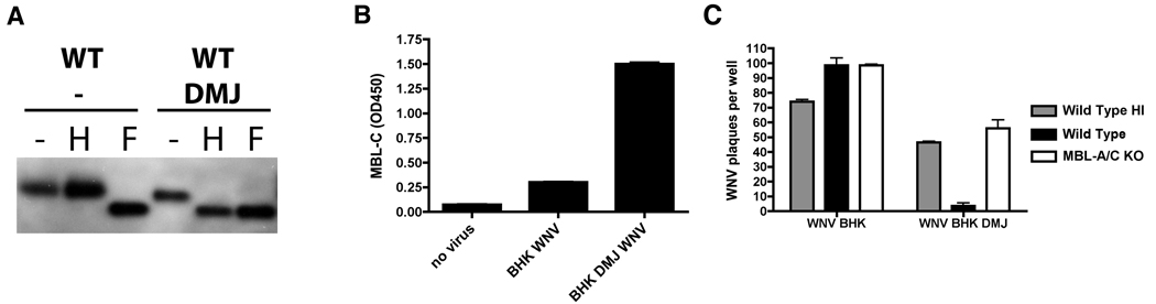
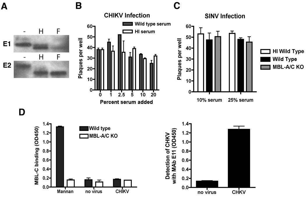
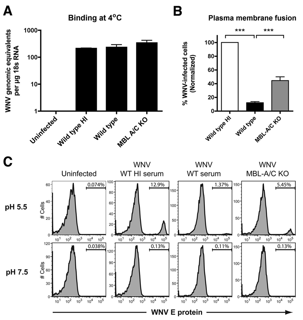
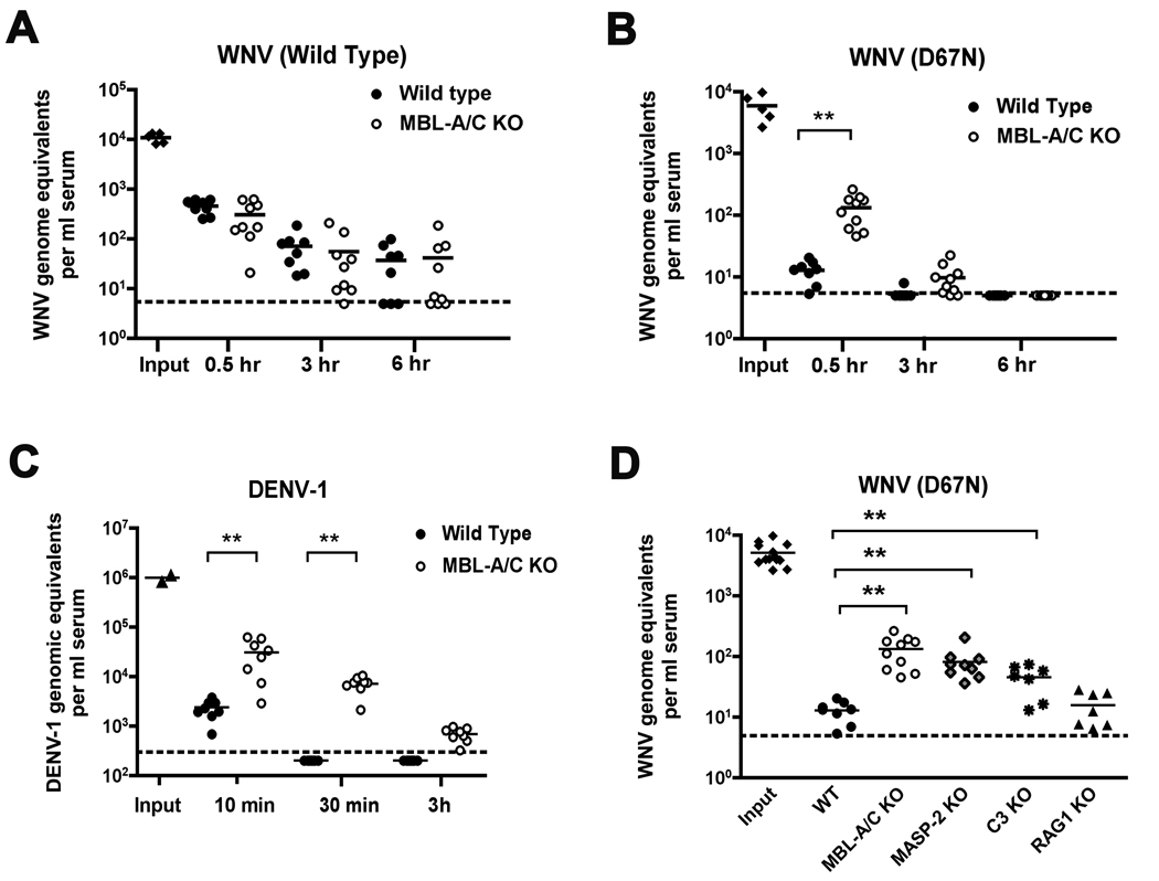
An intact complement system is crucial for limiting West Nile virus (WNV) dissemination. Herein, we define how complement directly restricts flavivirus infection in an antibody-independent fashion. Mannose-binding lectin (MBL) recognized N-linked glycans on the structural proteins of WNV and Dengue virus (DENV), resulting in neutralization through a C3- and C4-dependent mechanism that utilized both the canonical and bypass lectin activation pathways. For WNV, neutralization occurred with virus produced in insect cells, whereas for DENV, neutralization of insect and mammalian cell-derived virus was observed. Mechanism of action studies suggested that the MBL-dependent neutralization occurred, in part, by blocking viral fusion. Experiments in mice showed an MBL-dependent accelerated intravascular clearance of DENV or a WNV mutant with two N-linked glycans on its E protein, but not with wild-type WNV. Our studies show that MBL recognizes terminal mannose-containing carbohydrates on flaviviruses, resulting in neutralization and efficient clearance in vivo.


Figures

Figure 1
Figure 1. MBL binds to WNV virions and neutralizes infection in a MASP-2-and C3-dependent pathway
A–B. WNV virions were captured using an anti-WNV E protein-specific MAb and incubated with naïve serum from wild type (WT) (A, B) or MBL-A−/− x MBL-C−/− mice (MBL-A/C KO) (B). MBL-C binding to WNV was detected with an anti-MBL-C MAb. MBL-C binding was inhibited by the presence of 10 mM EDTA, 100 µg/ml mannan, or heat-inactivation (WT HI serum). The data are representative of two to three independent experiments performed in duplicate. C. WNV was pre-incubated with naïve serum from wild type, MBL-A−/− x MBL-C−/−, or RAG1 mice. Virus was added to BHK21 cells and the infectivity was determined three days later by plaque assay. The data are representative of two independent experiments performed in duplicate. D. The addition of recombinant MBL-C to serum from MBL-A−/− x MBL-C−/− mice restores neutralization of WNV. The data are representative of three independent experiments performed in duplicate. E. Neutralization of WNV in the presence of 10% (v/v) complement-deficient serum. Data are presented as percent neutralization (% reduction of plaque numbers over wild type heat-inactivated serum) by a given complement-deficient or antibody-deficient (RAG1) serum. Data are pooled from up to 15 independent experiments, and asterisks indicate values that are statistically different (P < 0.05) than that of wild type C57BL/6 serum. F. MBL-dependent neutralization does not promote virolysis. WNV was treated with media, detergent (NP-40, positive control), or serum from wild type (untreated or heat-inactivated) or MBL-A−/− x MBL-C−/− mice. RNase A was added, after which viral RNA was harvested and subjected to RT-PCR. The data is the average of four independent experiments and the dashed line indicates the limit of detection of the assay. G. C3 deposition on insect cell-derived WNV was measured in a capture ELISA using an anti-mouse C3 detection antibody and serum from various deficient mouse strains. One representative experiment of three is shown. H. Scheme of the lectin pathway of complement activation. MBL binding induces activation of C3 either via C4 and C2, or through a C2 bypass pathway. I. Mouse dendritic cells were infected with WNV that was pre-treated without serum, or with serum (untreated or heat-inactivated) from wild type or MBL-A−/− x MBL-C−/− mice. One day later virus was harvested from supernatants and titrated by plaque assay. The data are an average of three independent experiments performed in triplicate, and asterisks indicate values that are statistically different (P < 0.04). Error bars represent standard deviation (panels B, D–G, and I) or standard error of the mean (panels A and C).
Figure 2
Figure 2. MBL binds strongly to and neutralizes insect cell-derived but not mammalian cell-derived WNV
A. Western blot of E protein from WNV virions produced in (left) insect (C6/36) or (right) mammalian (BHK21) cells. To assess the type of N-linked glycan on WNV E proteins, samples were treated with Endo H (H) or PNGase F (F). B. MBL-C binding was assayed after WNV generated from insect or mammalian cells was incubated with serum from wild type or MBL-A−/− x MBL-C−/− mice as described in Fig 1. Equal virus loading of microtiter wells was confirmed by detection with an anti-WNV E protein MAb performed in parallel (data not shown). The data are an average of two independent experiments performed in duplicate. C. Insect or mammalian cell-derived WNV was incubated with untreated or heat-inactivated naive serum from wild type or MBL-A−/− x MBL-C−/− mice, and C3 deposition was measured by ELISA as described in Fig 1. The data are an average of three independent experiments performed in duplicate, and asterisks indicate values that are statistically different (P < 0.05) than that observed in the absence of virus. D. Serum neutralization of WNV derived from C6/36 or Vero cells. Experiments were performed as in Fig 1, with a final concentration of 10% of the indicated serum. The data are representative of two independent experiments performed in duplicate. Error bars represent standard deviation (panels B and C) or standard error of the mean (panel D).
Figure 3
Figure 3. MBL binds and neutralizes mammalian cell-derived WNV generated in the presence of the α-(1,2) mannosidase I inhibitor DMJ
WNV was generated in BHK21 cells in the presence or absence of DMJ. A. Western blot showing differences in E protein migration after treatment with (H) Endo H or (F) PNGase F of virus generated in BHK21 cells in the absence or presence of DMJ. The panel shows one of two experiments with identical results. B–C. MBL binding (B) and serum neutralization (C) of WNV from BHK21 cells that were untreated or treated with DMJ were tested as described in Fig 1. The data are representative of two independent experiments performed in duplicate. Error bars in this Figure represent standard deviations.
Figure 4
Figure 4. MBL-dependent neutralization of DENV
A. MBL binding and serum neutralization of DENV-2 were assessed using a virus capture ELISA (A) and plaque assay (B and C), respectively. DENV-2 was generated in insect cells for (A) MBL ELISA and (B) serum neutralization or in BHK21 mammalian cells (C) for neutralization. The data are an average of two to four independent experiments performed in duplicate. D. A mutant WNV was engineered with a “Dengue-like” glycosylation pattern in E (two N-linked glycan sites: D67N, N154) and was propagated in the presence or absence of DMJ in BHK21 cells. A Western blot with anti-E MAbs of virions after treatment with Endo H (H) or PNGase F (F) treatment is shown. Note, the D67N showed incomplete addition of N-linked glycans to the WNV E protein. As a control, D67N viral isolates were sequenced and showed complete preservation of the mutation. E–G. D67N WNV was propagated in mammalian or insect cells, and assessed for (E) MBL binding and (F and G) serum neutralization. The data are representative of two and four independent experiments, respectively, performed in duplicate. Error bars represent standard deviation (panels A, and E–G) or standard error of the mean (panels B and C).
Figure 5
Figure 5. MBL does not neutralize alphavirus infection
A. Western blot of CHIKV E1 and E2 proteins from insect cell passaged virus with and without treatment of (H) Endo H or (F) PNGase F. B–C. Neutralization of (B) CHIKV and (C) SINV was assessed in the presence or absence of wild type serum as described in the Fig 1. D. MBL binding to CHIKV was evaluated using a modified virus capture ELISA as described in Fig 1. As a positive control MBL-C in wild type serum bound to mannan (left column). However, MBL-C binding was not detected to either CHK Fab fragments alone or with CHIKV (middle and right columns). Detection with MAb E11 (anti-E2) antibody (right panel) demonstrates the level of virus addition in microtiter wells. The data are representative of two to three independent experiments performed in duplicate. Error bars in this Figure represent standard deviations.
Figure 6
Figure 6. MBL-mediated complement activation neutralizes WNV infectivity likely by blocking viral fusion
A. MBL binding to WNV does not inhibit viral attachment to host cells. WNV was pre-incubated with heat-inactivated (Wild Type HI) or untreated (Wild Type) serum from C57BL6 mice, or with serum from MBL-A−/− x MBL-C−/− mice. Serum-opsonized virus was cooled and added to BHK21 cells on ice. After extensive washing, attachment was assessed by qRT-PCR. The data are representative of three independent experiments performed in triplicate. No statistically significant difference was observed. B–C. WNV fusion at the plasma membrane is impaired following incubation with serum from wild type but not MBL-A−/− x MBL-C−/− mice. Serum-opsonized WNV was incubated with BHK21 cells to allow viral attachment. Viral fusion at the plasma membrane was induced after a brief exposure to a low pH (5.5) buffer. After pH neutralization, cells were cultured for 24 hrs in the presence of NH4Cl to inhibit viral infection through the endosomal pathway. Cells were analyzed for the presence of intracellular virus by staining with an anti-E protein MAb. Representative histograms are shown (C) and the data was pooled from four experiments performed for statistical analysis (B). Asterisks indicate values that are statistically different (P < 0.05). Note the efficiency of low pH-triggered viral fusion at the plasma membrane is much lower than that observed with endosomal fusion pathways. Error bars in this Figure represent standard deviations.
Figure 7
Figure 7. WNV D67N mutant and DENV are cleared from the circulation of mice with faster kinetics in an MBL-dependent manner
A–B. Clearance of insect-cell derived WNV in mice. Wild type or MBL-A−/− x MBL-C−/− mice were inoculated with 5 × 10⁴ PFU of (A) parent wild type or (B) D67N mutant WNV by an intravenous route. Blood was collected at 0.5, 3 and 6 hrs post-injection, and viral RNA in serum was measured. The results are from 8 to 10 mice per group, and asterisks indicate differences that are statistically significant (P < 0.05). C. Wild type and MBL-A−/− x MBL-C−/− mice were infected i.v. with 106 PFU of DENV-1. After harvesting blood at the indicated times, viral RNA in serum was measured. The results are from 8 to 9 mice per group, and asterisks indicate differences that are statistically significant (P < 0.05). D. Clearance of insect-cell derived WNV D67N in mice requires C3 activation but not antibody. Wild type, MBL-A−/− x MBL-C−/−, MASP-2−/−, C3−/−, and RAG1−/− mice were inoculated with 5 × 10⁴ PFU of WNV D67N by an intravenous route. Blood was collected at 0.5 hrs post-injection, and viral RNA in serum was measured. The results are from 7 to 10 mice per group, and asterisks indicate differences that are statistically significant (P < 0.05).

Similar articles

Cited by

References

    1. Acioli-Santos B, Segat L, Dhalia R, Brito CA, Braga-Neto UM, Marques ET, Crovella S. MBL2 gene polymorphisms protect against development of thrombocytopenia associated with severe dengue phenotype. Hum Immunol. 2008;69:122–128. - PubMed
    1. Avirutnan P, Mehlhop E, Diamond MS. Complement and its role in protection and pathogenesis of flavivirus infections. Vaccine. 2008;26 Suppl 8:I100–I107. - PMC - PubMed
    1. Bathum L, Hansen H, Teisner B, Koch C, Garred P, Rasmussen K, Wang P. Association between combined properdin and mannose-binding lectin deficiency and infection with Neisseria meningitidis. Mol Immunol. 2006;43:473–479. - PubMed
    1. Beasley DW, Whiteman MC, Zhang S, Huang CY, Schneider BS, Smith DR, Gromowski GD, Higgs S, Kinney RM, Barrett AD. Envelope protein glycosylation status influences mouse neuroinvasion phenotype of genetic lineage 1 West Nile virus strains. J Virol. 2005;79:8339–8347. - PMC - PubMed
    1. Bokisch VA, Top FH, Jr, Russell PK, Dixon FJ, Muller-Eberhard HJ. The potential pathogenic role of complement in dengue hemorrhagic shock syndrome. N Engl J Med. 1973;289:996–1000. - PubMed

Publication types

MeSH terms