. 2010 Dec;46(18):3365-74.
doi: 10.1016/j.ejca.2010.07.007. Epub 2010 Aug 13.

Molecular targets of apigenin in colorectal cancer cells: involvement of p21, NAG-1 and p53

Affiliations

Molecular targets of apigenin in colorectal cancer cells: involvement of p21, NAG-1 and p53

Yi Zhong et al. Eur J Cancer. 2010 Dec.

Abstract

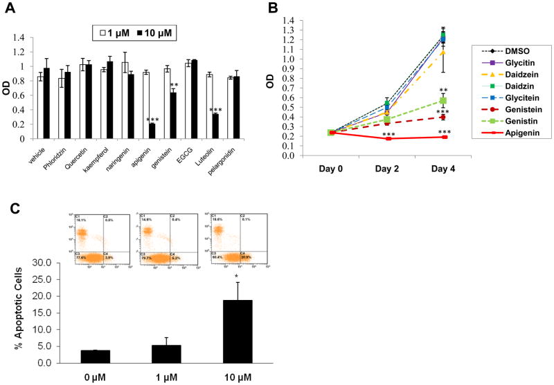
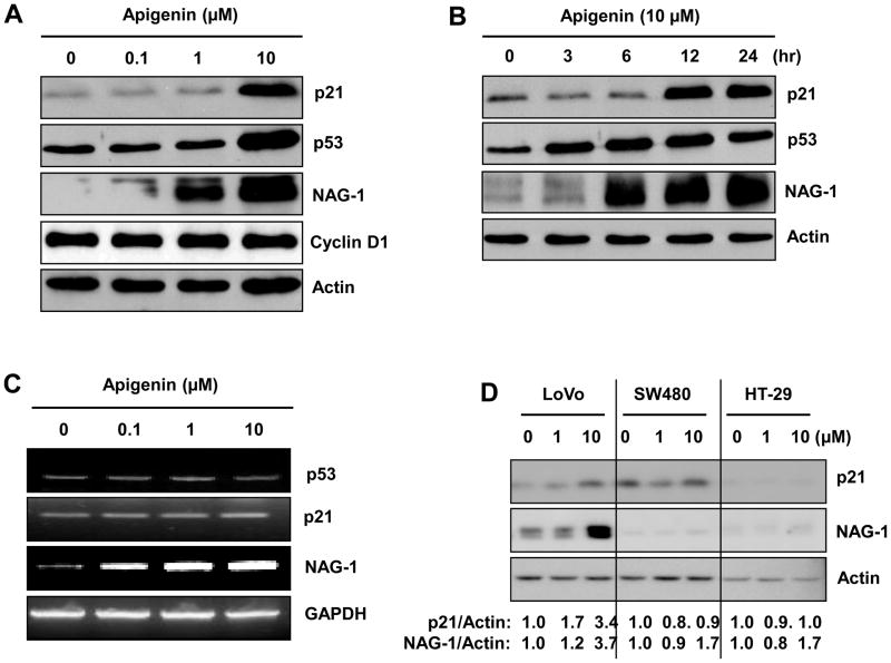
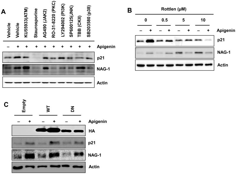
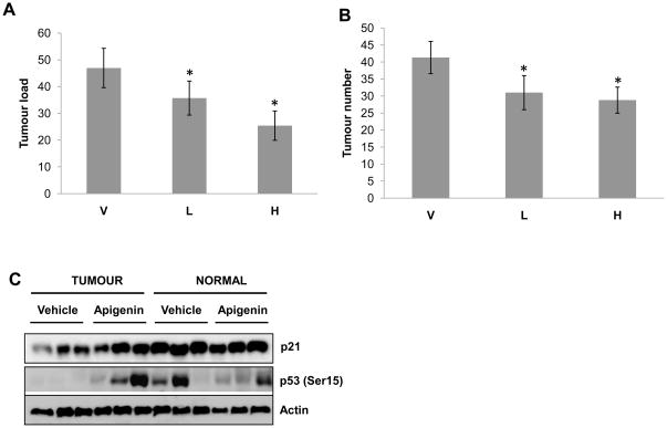
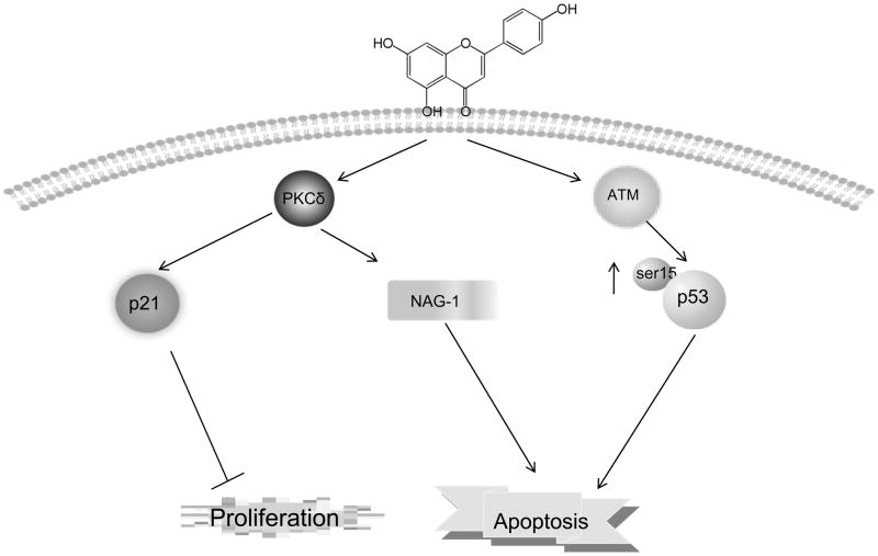
Persuasive epidemiological and experimental evidence suggests that dietary flavonoids have anti-cancer activity. Since conventional therapeutic and surgical approaches have not been able to fully control the incidence and outcome of most cancer types, including colorectal neoplasia, there is an urgent need to develop alternative approaches for the management of cancer. We sought to develop the best flavonoids for the inhibition of cell growth, and apigenin (flavone) proved to be the most promising compound in colorectal cancer cell growth arrest. Subsequently, we found that pro-apoptotic proteins (NAG-1 and p53) and cell cycle inhibitor (p21) were induced in the presence of apigenin, and kinase pathways, including PKCδ and ataxia telangiectasia mutated (ATM), play an important role in activating these proteins. The data generated by in vitro experiments were confirmed in an animal study using APC(MIN+) mice. Apigenin is able to reduce polyp numbers, accompanied by increasing p53 activation through phosphorylation in animal models. Our data suggest apparent beneficial effects of apigenin on colon cancer.


Conflict of interest statement

Conflict of Interest

No conflict of interest exists in the submission of this manuscript

Figures

Figure 1
Figure 1
Effect of apigenin and other flavonoids on colon cancer cell growth and apoptosis. A) HCT-116 cells were treated with DMSO or various compounds at concentrations of 1 μM and 10 μM for 4 days. Cell growth was measured using the Cell Titer96® Aqueous one solution cell proliferation assay as described in the Materials and Methods section. Values are expressed as mean±SD of five replicates. **p<0.01, ***p<0.001 versus DMSO-treated cells at day 4. B) HCT-116 cells were treated with DMSO or various flavones and isoflavones at 10 μM concentration for 0, 2, and 4 days. Cell growth was measured using the Cell Titer96 Aqueous one solution cell proliferation assay. **p<0.01,***p<0.001 versus DMSO-treated cells at day 4. C) HCT-116 cells were plated at 5×10⁵cells/well in six-well plates, incubated with 1 μM and 10 μM of apigenin for 72 h and analyzed for apoptosis as described in Materials and Methods. Values are expressed as mean±SD of triplicates.*p<0.5 versus DMSO-treated cells.
Figure 2
Figure 2
Analysis of p21, p53 and NAG-1 expressions in colorectal cancer cells treated with apigenin. A) Western blot analysis of p21, p53, cyclinD1, NAG-1 and actin expression in HCT-116 cells treated with indicated dose of apigenin for 24 h. B) HCT-116 cells were treated with 10 μM of apigenin, and samples were harvested at different time points. Cell lysates were then subjected to Western blot analysis for p21, p53, NAG-1 and actin. C) HCT-116 cells were treated with 0, 0.1 μM, 1 μM, and 10 μM apigenin and then RT-PCR was performed. Number of cycles used was 29 for p53, p21 and NAG-1, and 25 cycles for GAPDH. Reaction products were analyzed on 2% agarose gel. D) Western blot analysis of p21, NAG-1 and actin in different colon cancer cells treated with 0, 1, and 10 μM of apigenin for 24 h. Relative densitometry is shown in the bottom of the figure, as vehicle-treated sample is 1.0.
Figure 3
Figure 3
Apigenin effects on p53 expression. Western blot analysis of phosphor-p53 Ser-15, Ser-37, Ser-6, Ser-392 and actin in HCT-116 cells. A) HCT-116 cells were treated with 0, 0.1 μM, 1 μM and 10 μM of apigenin for 24 h. B) HCT-116 cells were treated with 10 μM apigenin with different time points. C) Apigenin increased p53-dependent transcriptional activity. The pMdm-2 construct was transiently transfected into HCT-116 cells. After 24 h transfection, cells were treated with either DMSO or 10 μM apigenin for 24 h, and luciferase activity was measured. The value represents mean±SD from three independent experiments. D) HCT-116 p53−/− cells were treated with 0.1 μM, 1 μM, and 10 μM apigenin for 24 h, and Western blot analysis was performed using antibodies for p21, NAG-1 and actin.
Figure 4
Figure 4
Effects of kinase inhibitors on apigenin-induced p21, and NAG-1 expression. A) HCT-116 cells were pretreated with DMSO, ATM inhibitor (KU55933, 10 μM), p38MAPK inhibitor (SB203580, 10 μM), CKII inhibitor (TBB, 5 μM), JNK inhibitor (SP600125, 10 μM), PI3K inhibitor (LY294002, 20 μM), PKC inhibitor (RO-318220, 2.5 μM), JAK-2 inhibitor (AG490, 50 μM), and pan-kinase inhibitor (staurosporine, 5μM), for 30 min and followed by treatment with 10 μM of apigenin for an additional 24 h. The cell lysates were harvested and subjected to Western blot anaylsis to examine p21, NAG-1, and actin expression. B) HCT-116 cells were treated with DMSO, and different concentrations of Rottlerin (0.5, 5, 10 μM) in serum-free media. After 30 min pre-treatment, either DMSO or 10 μM of apigenin was added directly to the media. After 24 h incubation, cell lysates were harvested and then subjected to Western blot using antibodies for p21, NAG-1, and actin. C) HCT-116 cells were transfected with empty, wild type (WT), or dominant negative (DN) PKCδ expression vector as described previously (43). The cells were then treated with 10 μM of apigenin for 24 h. Western analysis was performed for hemagglutinin (HA), NAG-1, p21, and actin antibodies.
Figure 5
Figure 5
Apigenin effect on APCMIN+ mice. (A and B) Effects of administration of apigenin or 0.5% methylcellulose (V) on tumour number and tumour load (mean ± SE) in comparison to vehicle (n=6), low dosage (L) of 25 mg/kg apigenin (n=6), and high dosage (H) of 50 mg/kg (n=5). Tumour load was calculated by number of tumors × average diameter. Tumour load and tumour numbers were statistically analyzed using the Mixed ANOVA test with statistical significance set at *p<0.05, compared to vehicle-treated mice. C) Cell lysates from tissue samples of three tumour and three normal tissues were isolated from vehicle-treated and apigenin-treated APCMIN+ mice (high dosage 50 mg/kg). Western blot anaylsis was performed for p21, phosphor-p53 (Ser-15), and actin.
Figure 6
Figure 6
Mechanism by which apigenin induces apoptosis and suppresses cell growth in HCT-116 cells. Apigenin affects the PKCδ pathway to increase p21 and NAG-1 expressions, followed by inducing apoptosis and inhibiting cell proliferation. Phosphor-p53 at Ser-37 and Ser-15, which are active forms, were increased by apigenin through the ATM pathway. Overall, apigenin affects p21 and NAG-1 expression in a p53- independent pathway.

Similar articles

Cited by

References

    1. Jemal A, Siegel R, Ward E, Hao Y, Xu J, Murray T, et al. Cancer statistics, 2008. CA Cancer J Clin. 2008;58(2):71–96. - PubMed
    1. Llorens F, Miró FA, Casañas A, Roher N, Garcia L, Plana M, et al. Unbalanced activation of ERK1/2 and MEK1/2 in apigenin-induced HeLa cell death. Experimental Cell Research. 2004;299(1):15–26. - PubMed
    1. Agullo G, Gamet-Payrastre L, Manenti S, Viala C, Rémésy C, Chap H, et al. Relationship between flavonoid structure and inhibition of phosphatidylinositol 3-kinase: A comparison with tyrosine kinase and protein kinase C inhibition. Biochemical Pharmacology. 1997;53(11):1649–1657. - PubMed
    1. Wang W, Heideman L, Chung CS, Pelling JC, Koehler KJ, Birt DF. Cell-cycle arrest at G2/M and growth inhibition by apigenin in human colon carcinoma cell lines. Mol Carcinog. 2000;28(2):102–10. - PubMed
    1. Au A, Li B, Wang W, Roy H, Koehler K, Birt D. Effect of dietary apigenin on colonic ornithine decarboxylase activity, aberrant crypt foci formation, and tumorigenesis in different experimental models. Nutr Cancer. 2006;54(2):243–51. - PubMed

Publication types

MeSH terms