. 2010 Nov;299(5):L639-51.
doi: 10.1152/ajplung.00405.2009. Epub 2010 Aug 13.

High-molecular-weight hyaluronan is a novel inhibitor of pulmonary vascular leakiness

Affiliations

High-molecular-weight hyaluronan is a novel inhibitor of pulmonary vascular leakiness

Patrick A Singleton et al. Am J Physiol Lung Cell Mol Physiol. 2010 Nov.

Abstract

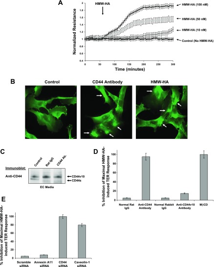
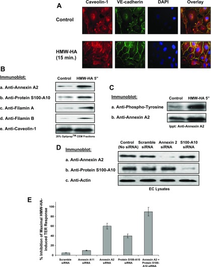
Endothelial cell (EC) barrier dysfunction results in increased vascular permeability, a perturbation observed in inflammatory states, tumor angiogenesis, atherosclerosis, and both sepsis and acute lung injury. Therefore, agents that enhance EC barrier integrity have important therapeutic implications. We observed that binding of high-molecular-weight hyaluronan (HMW-HA) to its cognate receptor CD44 within caveolin-enriched microdomains (CEM) enhances human pulmonary EC barrier function. Immunocytochemical analysis indicated that HMW-HA promotes redistribution of a significant population of CEM to areas of cell-cell contact. Quantitative proteomic analysis of CEM isolated from human EC demonstrated HMW-HA-mediated recruitment of cytoskeletal regulatory proteins (annexin A2, protein S100-A10, and filamin A/B). Inhibition of CEM formation [caveolin-1 small interfering RNA (siRNA) and cholesterol depletion] or silencing (siRNA) of CD44, annexin A2, protein S100-A10, or filamin A/B expression abolished HMW-HA-induced actin cytoskeletal reorganization and EC barrier enhancement. To confirm our in vitro results in an in vivo model of inflammatory lung injury with vascular hyperpermeability, we observed that the protective effects of HMW-HA on LPS-induced pulmonary vascular leakiness were blocked in caveolin-1 knockout mice. Furthermore, targeted inhibition of CD44 expression in the mouse pulmonary vasculature significantly reduced HMW-HA-mediated protection from LPS-induced hyperpermeability. These data suggest that HMW-HA, via CD44-mediated CEM signaling events, represents a potentially useful therapeutic agent for syndromes of increased vascular permeability.


Figures

Fig. 1.
Fig. 1.
Analysis of CD44 and caveolin-1 regulation of high-molecular-weight hyaluronan (HMW-HA) binding to human pulmonary endothelial cells (EC). A: EC were grown to confluency and serum-starved for 1 h, and Triton X-100-soluble, Triton X-100-insoluble, and OptiPrep fractions were prepared. The 20% OptiPrep fraction represents the caveolin-enriched microdomain (CEM) fraction. Fractions were run on SDS-PAGE, transferred to nitrocellulose, and immunoblotted with anti-caveolin-1 (a), anti-fibrillarin (b), anti-cyclooxygenase (COX) IV (c), anti-lysosomal-associated membrane glycoprotein 2 precursor (LAMP2b, d), anti-Golgi reassembly stacking protein 65 (GRASP65, e), or anti-VEGF receptor (anti-VEGFR, f). B: EC were grown to confluency, serum-starved for 1 h, and either left untreated (control) or treated with 100 nM HMW-HA (5 min) or the CEM-abolishing cholesterol-depletion agent methyl-β-cyclodextrin (MβCD, 5 mM) for 1 h prior to 100 nM HMW-HA treatment (5 min). Cellular material was solublized in 4°C 1% Triton X-100, and soluble and insoluble fractions were obtained. Triton X-100-insoluble fraction was overlaid with 60%, 40%, 30%, and 20% OptiPrep and centrifuged at 35,000 rpm in an SW60 rotor for 12 h at 4°C. Triton X-100-soluble material and OptiPrep fractions were run on SDS-PAGE, transferred to nitrocellulose, and immunoblotted with anti-caveolin-1 (a), anti-CD44s (IM-7, standard domain, b), or anti-VEGF receptor 2 (anti-VEGFR2, c) antibody. The 20% OptiPrep fraction is the CEM fraction. Experiments were performed in triplicate, with highly reproducible findings, and representative data are shown. C: immunoblot analysis of small interfering RNA (siRNA)-treated or untreated human EC. Cellular lysates from untransfected (control, no siRNA), scramble siRNA (siRNA that does not target any known human mRNA), caveolin-1 siRNA, or CD44 siRNA transfection were analyzed using immunoblotting with anti-caveolin-1 (a), anti-CD44 (IM-7, b), or anti-actin (c) antibody. Experiments were performed in triplicate, each with similar results, and representative data are shown. D: quantitation of fluorescein-conjugated HMW-HA binding to scramble siRNA-, annexin A11 siRNA-, CD44 siRNA-, or caveolin-1 siRNA-treated EC. Fluorescein-conjugated HMW-HA (100 nM) was added for 15 min to EC in serum-free medium, cells were washed 3 times in serum-free medium, and fluorescence intensity was quantified. Cells were counted utilizing a hemocytometer.
Fig. 2.
Fig. 2.
Analysis of CD44 and caveolin-1 regulation of HMW-HA-mediated human EC barrier enhancement. A: EC were plated on gold microelectrodes, serum-starved for 1 h, and treated with PBS, pH 7.4 (control), or 10, 50, or 100 nM HMW-HA. Arrow indicates HMW-HA addition. Transendothelial electrical resistance (TER) trace represents pooled means ± SE from 3 independent experiments. B: HMW-HA induces CD44 receptor clustering. Human pulmonary microvascular EC were plated on 8-well glass cover slides and allowed to adhere for 48 h. Cells were then serum-starved for 1 h and incubated with 100 nM HMW-HA for 15 min and fixed in 4% paraformaldehyde or incubated with rat anti-CD44 (IM-7) antibody at 10 μg/ml for 2 h. Cells were then washed briefly in PBS, pH 7.4, and secondary anti-rat IgG (1 μg/ml; Sigma-Aldrich) was added for CD44 antibody cross-linking (36). Cells were incubated for 1 h before fixation in 4% paraformaldehyde. Cells were then immunostained for CD44 with use of a directly labeled mouse anti-CD44-Alexa 488 conjugate (Cell Signaling Technology). C: anti-CD44 antibody (IM-7) does not induce appreciable CD44 shedding in human EC. Human EC were serum-starved for 1 h prior to the addition of no antibody, rat IgG (1 μg/ml), or rat anti-CD44 (IM-7) antibody (1 μg/ml) for 3 h. EC medium was collected, concentrated, and immunoblotted with anti-CD44 (IM-7) antibody. D: percent inhibition of maximal HMW-HA-induced TER response (as described in A) with addition of normal rat IgG, anti-CD44 antibody, normal rabbit IgG, anti-CD44v10 antibody (10 μg/ml), or 5 mM MβCD. E: percent inhibition of maximal HMW-HA-induced TER response (as described in D) in human EC with scramble, annexin A11, CD44, or caveolin-1 siRNA treatment.
Fig. 3.
Fig. 3.
Role of HMW-HA-induced recruitment of annexin A2 and protein S100-A10 to human EC CEM. A: HMW-HA induces caveolin-1 redistribution to EC-EC junctions. Human EC were grown to confluency, serum-starved for 1 h, and either left untreated (control) or treated with 100 nM HMW-HA (15 min), fixed in 4% paraformaldehyde, and stained with anti-caveolin-1 antibody, anti-vascular endothelial (VE)-cadherin antibody, or 4′,6-diamidino-2-phenylindole (DAPI). Overlay is a merged image of caveolin-1, VE-cadherin, and DAPI fluorescence, with yellow color indicating colocalization of caveolin-1 and VE-cadherin. B and C: EC were grown to confluency, serum-starved for 1 h, and either left untreated (control) or treated with 100 nM HMW-HA (5 min) and CEM fractions (20% OptiPrep layer). B: CEM fractions were run on SDS-PAGE, transferred to nitrocellulose, and immunoblotted with anti-annexin A2 (a), anti-protein S100-A10 (b), anti-filamin A (c), anti-filamin B (d), or anti-caveolin-1 (e) antibody. Experiments were performed in triplicate, with highly reproducible findings, and representative data are shown. C: CEM fractions were solublized in immunoprecipitation (Ippt) buffer and immunoprecipitated with anti-annexin A2 antibody. Resulting immunobeads were run on SDS-PAGE, transferred to nitrocellulose, and immunoblotted with anti-phosphotyrosine (a) or anti-annexin A2 (b) antibody. Experiments were performed in triplicate, with highly reproducible findings, and representative data are shown. D: EC were treated with no siRNA (control), scramble siRNA, annexin A2 siRNA, or protein S100-A10 siRNA for 48 h. EC lysates were obtained and run on SDS-PAGE, transferred to nitrocellulose, and immunoblotted with anti-annexin A2 (a), anti-protein S100-A10 (b), or anti-actin (c) antibody. Experiments were performed in triplicate, with highly reproducible findings, and representative data are shown. E: percent inhibition of maximal HMW-HA-induced TER response in human EC with scramble, annexin A11, annexin A2, protein S100-A10, or annexin A2 + protein S100-A10 siRNA treatment. Silencing both annexin II and protein S100-A10 is required for maximal inhibition of HMW-HA-induced TER in EC.
Fig. 4.
Fig. 4.
Annexin A2 and protein S100-A10 regulation of HMW-HA-induced filamin A/B recruitment to CEM and human EC barrier enhancement. A: immunoblot analysis of HMW-HA-treated (100 nM, 5 min) or untreated human EC lysates from scramble siRNA, annexin A2 siRNA, protein S100-A10 siRNA, or annexin A2 + protein S100-A10 siRNA transfection using anti-filamin A (a), anti-filamin B (b), or anti-caveolin-1 (c) antibody. Experiments were performed in triplicate, each with similar results, and representative data are shown. Silencing annexin A2 and protein S100-A10 blocks HMW-HA-induced filamin A and filamin B recruitment to CEM. B: EC were treated with no siRNA (control), scramble siRNA, filamin A siRNA, or filamin B siRNA for 48 h. EC lysates were obtained and run on SDS-PAGE, transferred to nitrocellulose, and immunoblotted with anti-filamin A (a), anti-filamin B (b), or anti-actin (c) antibody. Experiments were performed in triplicate, with highly reproducible findings, and representative data are shown. C: percent inhibition of maximal HMW-HA-induced TER response. EC were plated on gold microelectrodes and treated with scramble siRNA (control), annexin A11 siRNA, filamin A siRNA, filamin B siRNA, or filamin A + filamin B siRNA for 48 h. After EC were serum-starved for 1 h, 100 nM HMW-HA was added. Values represent pooled TER data ± SE at 30 min after addition of agonist from 3 independent experiments.
Fig. 5.
Fig. 5.
Analysis of HMW-HA-mediated human EC actin cytoskeletal rearrangement. A: immunofluorescent images of HMW-HA-induced EC cortical actin rearrangement. EC were serum-starved for 1 h and either left untreated (control) or treated with 100 nM HMW-HA for 15 min or pretreated with 5 mM MβCD for 1 h and then with 100 nM HMW-HA for 15 min. Cells were then fixed and stained with tetramethylrhodamine isothiocyanate (TRITC)-phalloidin (to visualize F-actin) and analyzed using fluorescent microscopy. Observations are representative of the entire cell monolayer and were reproduced in multiple independent experiments (n ≥ 3 for each condition). B: immunofluorescent images of HMW-HA-induced EC cortical actin rearrangement as described in A. Human EC were treated with scramble siRNA (control), CD44 siRNA, caveolin-1 siRNA, annexin A2 siRNA, or protein S100-A10 siRNA for 48 h. After EC were serum-starved for 1 h, 100 nM HMW-HA was added for 15 min.
Fig. 6.
Fig. 6.
Pulmonary vascular CD44 regulation of HMW-HA-induced protection from LPS-induced vascular hypermeability. A: immunohistochemical fluorescently stained images of control (untreated) mouse lung [bright-field (DIC) imaging (a) or treatment with anti-Factor VIII (von Willebrand factor) antibody (b), anti-caveolin-1 antibody (c), or FITC-conjugated anti-CD44 antibody (d) and secondary fluorescent antibody (Alexa Fluor 610 for vWF and 350 for caveolin-1; e)]. Magnification ×100. Arrows in e (an overlay of b, c, and d) indicate immunostaining of endothelial cells. Insets: negative controls for immunohistochemical analysis, which were obtained by the method described above, but without primary antibody. B: bronchoalveolar lavage (BAL) protein concentration from wild-type (C57BL/6J), CD44 knockout, or caveolin-1 knockout mice that were anesthetized and given saline (control) or LPS (2.5 mg/kg) intratracheally. After 4 h, mice were injected intravenously (internal jugular vein) with saline (control) or HMW-HA (1.5 mg/kg). Treated mice were allowed to recover for 24 h, BAL fluids were obtained, and protein concentrations were determined. *Significant (P < 0.05) difference between LPS and HMW-HA + LPS (n = 6 per condition). C: immunohistochemical analysis of murine lungs and kidneys either left untreated or injected intravenously (internal jugular vein) with angiotensin I-converting enzyme (ACE) antibody-conjugated liposomes (DOTAP/DOPE) containing siControl siRNA (10 mg/kg) or siSTABLE CD44 siRNA (10 mg/kg) for 5 days. Vascular CD44 expression is inhibited in lung, but not kidney. D: immunocytochemical analysis of cytospin material from BAL of mice injected intravenously (internal jugular vein) with ACE antibody-conjugated liposomes (DOTAP/DOPE) containing siControl siRNA (10 mg/kg) or siSTABLE CD44 siRNA (10 mg/kg) for 5 days. There is no difference in CD44 immunoreactivity. E: total BAL protein of B6129N2 mice injected intravenously (internal jugular vein) with ACE antibody-conjugated liposomes (DOTAP/DOPE) containing siControl siRNA (5 or 10 mg/kg) or siSTABLE CD44 siRNA (5 or 10 mg/kg) for 5 days (n = 6 per condition). *Statistically significant difference (P < 0.05) between siControl and siCD44. F: BAL protein concentration from C57BL/6J mice injected intravenously (internal jugular vein) with ACE antibody-conjugated liposomes (DOTAP/DOPE) containing scramble siRNA (10 mg/kg) or siSTABLE CD44 siRNA (10 mg/kg) for 5 days. Mice were then anesthetized and given saline (control) or LPS (2.5 mg/kg) intratracheally. After 4 h, mice were injected intravenously (internal jugular vein) with saline (control) or HMW-HA (1.5 mg/kg). Treated mice were allowed to recover for 24 h, BAL fluids were obtained, and protein concentrations were determined. *Significant (P < 0.05) difference between LPS and HMW-HA + LPS (n = 6 per condition).
Fig. 7.
Fig. 7.
Proposed model of HMW-HA-induced vascular integrity. HMW-HA binding to CD44s in caveolin-enriched microdomains (CEM) in human EC (1) induces annexin A2 tyrosine phosphorylation and annexin A2/protein S100-A10 translocation to CEM (2). Annexin A2 and protein S100-A10 are crucial for subsequent HMW-HA-induced recruitment of filamin A and filamin B to CEM (3), actin cytoskeletal reorganization (cortical actin formation) (4), and EC barrier enhancement (5).

References

    1. Balyasnikova IV, Sun ZL, Metzger R, Taylor PR, Vicini E, Muciaccia B, Visintine DJ, Berestetskaya YV, McDonald TD, Danilov SM. Monoclonal antibodies to native mouse angiotensin-converting enzyme (CD143): ACE expression quantification, lung endothelial cell targeting and gene delivery. Tissue Antigens 67: 10–29, 2006. - PubMed
    1. Bensadoun ES, Burke AK, Hogg JC, Roberts CR. Proteoglycan deposition in pulmonary fibrosis. Am J Respir Crit Care Med 154: 1819–1828, 1996. - PubMed
    1. Borbiev T, Verin AD, Shi S, Liu F, Garcia JG. Regulation of endothelial cell barrier function by calcium/calmodulin-dependent protein kinase II. Am J Physiol Lung Cell Mol Physiol 280: L983–L990, 2001. - PubMed
    1. Bourguignon LY, Gilad E, Brightman A, Diedrich F, Singleton P. Hyaluronan-CD44 interaction with leukemia-associated RhoGEF and epidermal growth factor receptor promotes Rho/Ras co-activation, phospholipase Cε-Ca2+ signaling, and cytoskeleton modification in head and neck squamous cell carcinoma cells. J Biol Chem 281: 14026–14040, 2006. - PubMed
    1. Bourguignon LY, Ramez M, Gilad E, Singleton PA, Man MQ, Crumrine DA, Elias PM, Feingold KR. Hyaluronan-CD44 interaction stimulates keratinocyte differentiation, lamellar body formation/secretion, and permeability barrier homeostasis. J Invest Dermatol 126: 1356–1365, 2006. - PubMed

Publication types

MeSH terms

LinkOut - more resources