. 2010 Dec;299(6):L735-48.
doi: 10.1152/ajplung.00153.2010. Epub 2010 Aug 13.

Moderate postnatal hyperoxia accelerates lung growth and attenuates pulmonary hypertension in infant rats after exposure to intra-amniotic endotoxin

Affiliations

Moderate postnatal hyperoxia accelerates lung growth and attenuates pulmonary hypertension in infant rats after exposure to intra-amniotic endotoxin

Jen-Ruey Tang et al. Am J Physiol Lung Cell Mol Physiol. 2010 Dec.

Abstract

To determine the separate and interactive effects of fetal inflammation and neonatal hyperoxia on the developing lung, we hypothesized that: 1) antenatal endotoxin (ETX) causes sustained abnormalities of infant lung structure; and 2) postnatal hyperoxia augments the adverse effects of antenatal ETX on infant lung growth. Escherichia coli ETX or saline (SA) was injected into amniotic sacs in pregnant Sprague-Dawley rats at 20 days of gestation. Pups were delivered 2 days later and raised in room air (RA) or moderate hyperoxia (O₂, 80% O₂ at Denver's altitude, ∼65% O₂ at sea level) from birth through 14 days of age. Heart and lung tissues were harvested for measurements. Intra-amniotic ETX caused right ventricular hypertrophy (RVH) and decreased lung vascular endothelial growth factor (VEGF) and VEGF receptor-2 (VEGFR-2) protein contents at birth. In ETX-exposed rats (ETX-RA), alveolarization and vessel density were decreased, pulmonary vascular wall thickness percentage was increased, and RVH was persistent throughout the study period compared with controls (SA-RA). After antenatal ETX, moderate hyperoxia increased lung VEGF and VEGFR-2 protein contents in ETX-O₂ rats and improved their alveolar and vascular structure and RVH compared with ETX-RA rats. In contrast, severe hyperoxia (≥95% O₂ at Denver's altitude) further reduced lung vessel density after intra-amniotic ETX exposure. We conclude that intra-amniotic ETX induces fetal pulmonary hypertension and causes persistent abnormalities of lung structure with sustained pulmonary hypertension in infant rats. Moreover, moderate postnatal hyperoxia after antenatal ETX restores lung growth and prevents pulmonary hypertension during infancy.


Figures

Fig. 1.
Fig. 1.
Effects of intra-amniotic antenatal endotoxin (ETX) and moderate postnatal hyperoxia on survival rate of infant rats. As shown, survival in the ETX-RA group was worse than SA-RA controls (P < 0.05 ETX-RA vs. SA-RA). SA-RA, saline-room air; ETX-RA, endotoxin-room air; SA-O2, saline-80% O2; ETX-O2, endotoxin-80% O2.
Fig. 2.
Fig. 2.
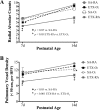
A–C: effects of intra-amniotic ETX and moderate postnatal hyperoxia on body weight of infant rats. As shown, body weight was decreased by ETX compared with saline controls from birth through 14 days of age (P < 0.05, ETX-RA vs. SA-RA), and ETX-O2 rats had higher body weights than ETX-RA rats at and after day 2 (P < 0.05).
Fig. 3.
Fig. 3.
Effects of intra-amniotic ETX and moderate postnatal hyperoxia on distal lung growth at days 7 and 14. A and C: lung micrographs are representative for each group and were obtained at the same magnification. Internal scale bar, 100 μm. B: at day 7, radial alveolar counts (RAC) were decreased in ETX-RA rats compared with SA-RA controls (P < 0.05), and ETX-O2 rats had higher RAC than ETX-RA rats (P < 0.05). D: at day 14, RAC remained decreased in ETX-RA rats compared with SA-RA controls and with ETX-O2 rats (P < 0.001 for each comparison).
Fig. 4.
Fig. 4.
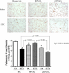
Effects of intra-amniotic ETX and moderate postnatal hyperoxia on pulmonary vessel density. A and C: representative findings are shown for each group at days 7 and 14 after immunostaining for von Willebrand Factor (vWF). Micrographs were obtained at the same magnification. Internal scale bar, 100 μm. B: at day 7, pulmonary vessel density was decreased in ETX-RA rats compared with SA-RA controls and ETX-O2 (P < 0.001 for each comparison). D: at day 14, pulmonary vessel density in ETX-RA rats remained decreased when compared with SA-RA controls and ETX-O2 (P < 0.001 for each comparison).
Fig. 5.
Fig. 5.
A and B: effects of intra-amniotic ETX and moderate postnatal hyperoxia on pulmonary vascular wall thickness from 14-day-old rats. Lung micrographs are representative and were obtained at the same magnification. Internal scale bar, 100 μm. Pulmonary vascular wall thickness percentage was increased in ETX-RA rats compared with SA-RA controls and ETX-O2 rats (P < 0.001 for each comparison).
Fig. 6.
Fig. 6.
Changes in RAC and pulmonary vessel density from postnatal day 7 to day 14. A: RAC were decreased in ETX-RA rats at day 7 (P < 0.05) and remained decreased at day 14 (P < 0.05) compared with SA-RA controls and ETX-O2 rats (P < 0.05 for each comparison). B: pulmonary vessel density was decreased in ETX-RA rats at day 7 (P < 0.05) and remained decreased at day 14 (P < 0.05) compared with SA-RA controls. ETX-O2 rats had higher vessel density than ETX-RA rats at days 7 and 14 (P < 0.001 for each age).
Fig. 7.
Fig. 7.
Effects of intra-amniotic ETX and moderate postnatal hyperoxia on the ratio of right ventricle to left ventricle plus septum weights (RV/LV+S) in infant rats. The RV/LV+S ratio was increased in ETX rats at birth compared with saline controls (P < 0.05; A) and remained elevated in ETX-RA rats throughout the study period (P < 0.05 for each age; B). Moderate hyperoxia reduced RV/LV+S ratio in ETX-O2 rats by day 2 compared with ETX-RA rats (P < 0.001).
Fig. 8.
Fig. 8.
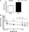
Effects of intra-amniotic ETX on oxygen saturation (SatO2) of infant rats in room air. SatO2 was decreased in ETX-RA newborn rats throughout 8 h of age compared with SA-RA controls (P < 0.05). P < 0.05 vs. SA-RA.
Fig. 9.
Fig. 9.
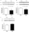
A–C: effects of intra-amniotic ETX on lung protein contents of vascular endothelial growth factor (VEGF), VEGF receptor-2 (VEGFR-2), and endothelial nitric oxide synthase (eNOS) at birth. Lung VEGF and VEGFR-2 protein contents were decreased in ETX rats compared with saline controls (P < 0.01 for VEGF; P < 0.05 for VEGFR-2). Lung eNOS protein content in ETX rats was comparable to saline controls at birth. There were 4–5 animals analyzed in each group.
Fig. 10.
Fig. 10.
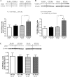
Effects of intra-amniotic ETX and moderate postnatal hyperoxia on lung protein contents of VEGF, VEGFR-2, and eNOS at postnatal day 5. P < 0.01 vs. SA-RA. P < 0.05 ETX-O2 vs. ETX-RA.
Fig. 11.
Fig. 11.
Comparison of the effects of 80% O2 and ≥95% O2 after intra-amniotic ETX on distal air space structure in 14-day-old rats. Lung micrographs are representative for each group and were obtained at the same magnification. There were 5–8 animals analyzed in each group. P < 0.05 vs. SA-RA.
Fig. 12.
Fig. 12.
Comparison of 80% O2 and ≥95% O2 effects after intra-amniotic ETX on pulmonary vessel density. Lung histology was stained with vWF, and micrographs are representative for each group. There were 5–8 animals analyzed in each group. P < 0.05 vs. SA-RA. HPF, high-powered field.

References

    1. Aggarwal NR, D'Alessio FR, Tsushima K, Files DC, Damarla M, Sidhaye VK, Fraig MM, Polotsky VY, King LS. Moderate oxygen augments lipopolysaccharide-induced lung injury in mice. Am J Physiol Lung Cell Mol Physiol 298: L371–L381, 2010 - PMC - PubMed
    1. Alvira CM, Abate A, Yang G, Dennery PA, Rabinovitch M. Nuclear factor-kappaB activation in neonatal mouse lung protects against lipopolysaccharide-induced inflammation. Am J Respir Crit Care Med 175: 805–815, 2007 - PMC - PubMed
    1. Andrews WW, Goldenberg RL, Faye-Petersen O, Cliver S, Goepfert AR, Hauth JC. The Alabama Preterm Birth study: polymorphonuclear and mononuclear cell placental infiltrations, other markers of inflammation, and outcomes in 23- to 32-wk preterm newborn infants. Am J Obstet Gynecol 195: 803–808, 2006 - PubMed
    1. Balasubramaniam V, Maxey AM, Morgan DB, Markham NE, Abman SH. Inhaled NO restores lung structure in eNOS-deficient mice recovering from neonatal hypoxia. Am J Physiol Lung Cell Mol Physiol 291: L119–L127, 2006 - PubMed
    1. Bancalari E, Claure N. Evolving clinical features of bronchopulmonary dysplasia. In: Bronchopulmonary Dysplasia. New York: Informa Health Care, 2010, p.208–222

Publication types

MeSH terms