. 2010 Oct;9(10):1418-31.
doi: 10.1128/EC.00097-10. Epub 2010 Aug 13.

The Saccharomyces cerevisiae anaphase-promoting complex interacts with multiple histone-modifying enzymes to regulate cell cycle progression

Affiliations

The Saccharomyces cerevisiae anaphase-promoting complex interacts with multiple histone-modifying enzymes to regulate cell cycle progression

Emma L Turner et al. Eukaryot Cell. 2010 Oct.

Abstract

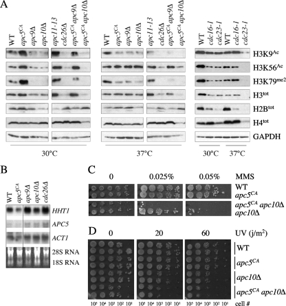
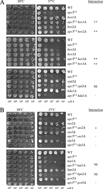
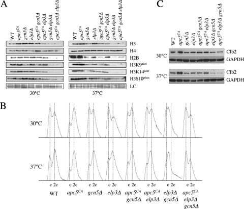
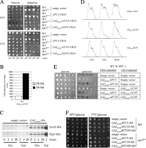
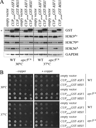
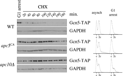
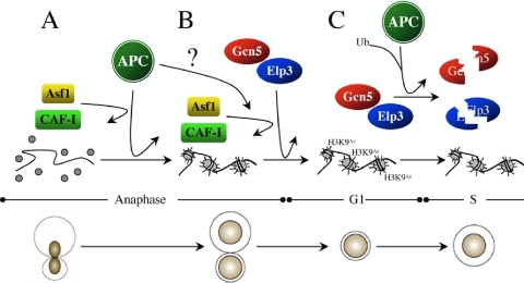
The anaphase-promoting complex (APC), a large evolutionarily conserved ubiquitin ligase complex, regulates cell cycle progression through mitosis and G(1). Here, we present data suggesting that APC-dependent cell cycle progression relies on a specific set of posttranslational histone-modifying enzymes. Multiple APC subunit mutants were impaired in total and modified histone H3 protein content. Acetylated H3K56 (H3K56(Ac)) levels were as reduced as those of total H3, indicating that loading histones with H3K56(Ac) is unaffected in APC mutants. However, under restrictive conditions, H3K9(Ac) and dimethylated H3K79 (H3K79(me2)) levels were more greatly reduced than those of total H3. In a screen for histone acetyltransferase (HAT) and histone deacetylase (HDAC) mutants that genetically interact with the apc5(CA) (chromatin assembly) mutant, we found that deletion of GCN5 or ELP3 severely hampered apc5(CA) temperature-sensitive (ts) growth. Further analyses showed that (i) the elp3Δ gcn5Δ double mutant ts defect was epistatic to that observed in apc5(CA) cells; (ii) gcn5Δ and elp3Δ mutants accumulate in mitosis; and (iii) turnover of the APC substrate Clb2 is not impaired in elp3Δ gcn5Δ cells. Increased expression of ELP3 and GCN5, as well as genes encoding the HAT Rtt109 and the chromatin assembly factors Msi1 and Asf1, suppressed apc5(CA) defects, while increased APC5 expression partially suppressed elp3Δ gcn5Δ growth defects. Finally, we demonstrate that Gcn5 is unstable during G(1) and following G(1) arrest and is stabilized in APC mutants. We present our working model in which Elp3/Gcn5 and the APC work together to facilitate passage through mitosis and G(1). To progress into S, we propose that at least Gcn5 must then be targeted for degradation in an APC-dependent fashion.


Figures

Fig. 1.
Fig. 1.
The APC is required for maintenance of total and modified histone H3 levels. (A) Extracts prepared from the APC mutants shown at the top of the lanes, grown at 30°C or after a 3-h shift to 37°C, were analyzed by Western analysis with the antibodies indicated on the right. Antibodies against GAPDH were used to control for protein load. (B) A Northern analysis was performed on the strains shown grown overnight at room temperature to early log phase. PCR fragments corresponding to the HHT1, APC5, and ACT1 ORFs were prepared, randomly labeled using [α-32P]CTP, and hybridized to purified and separated total RNA. (C and D) The mutants shown were grown overnight to early log phase in rich glucose media at 30°C. The next day, a 10-fold dilution series was prepared and spotted onto plates containing increasing concentrations of methyl methanesulfonate (MMS) (C) or plated onto YPD plates and exposed to an increasing UV dose (D). The plates exposed to UV were wrapped in aluminum foil and grown in the dark for 3 days. The approximate number of cells in each diluted spot is indicated below the plates.
Fig. 2.
Fig. 2.
Genetic interactions of HDAC and HAT mutants with the apc5CA allele. (A) apc5CA cells were repeatedly crossed with the mutants shown to generate double mutants. Tenfold serial dilutions of the different mutants were spot diluted onto rich media, and growth levels were compared at the temperatures shown. Plates were grown from 3 to 5 days and scanned each day. (B) apc5CA cells were repeatedly crossed with the mutants shown to generate double mutants. The mutants were prepared and analyzed as described above. The effect of the interaction is indicated on the right. ++, strong suppressive interaction in double mutant that grows like wild type; +, double mutant grows better than single mutants but not at wild-type level. −, double mutant grows more poorly than the single mutants. NI, no interaction.
Fig. 3.
Fig. 3.
Genetic interactions between apc5CA and HAT/HDAC mutants are complex. (A) A genetic interaction between hpa2Δ and gcn5Δ mutants is revealed in apc5CA cells. The mutants shown were constructed by genetic crosses and tested for temperature sensitivity using spot dilutions. See the legend to Fig. 2 for explanation of interaction symbols. (B) The elp3Δ gcn5Δ defect is more severe than that of strains containing apc5CA, as determined by spot dilutions. (C) The mutants shown were created through multiple rounds of backcrossing and characterized using spot dilutions. (D) Increased expression of APC5 partially suppresses elp3Δ gcn5Δ ts growth. WT and elp3Δ gcn5Δ cells were transformed with an APC5-expressing construct under the control of the GAL1 promoter or the empty vector control. The transformants were spot diluted onto glucose- or galactose-supplemented plates and grown for 3 to 13 days.
Fig. 4.
Fig. 4.
Histone posttranslational modifications and cell cycle profiles in apc5CA, elp3Δ, and gcn5Δ single, double, and triple mutants. (A) apc5CA, elp3Δ, and gcn5Δ mutants were used to characterize histone profiles. Extracts were prepared after growth at 30°C or following a shift to 37°C for 3 h. Proteins were separated by SDS-PAGE, and Western analysis was performed using the antibodies indicated. (B) Flow cytometry was conducted on the asynchronous cultures described for panel A. (C) Clb2 stability was assessed in the cells described for panel A. Cell cultures were prepared and proteins were extracted as described for panel A. Western analyses were performed using antibodies against endogenous Clb2 and GAPDH as a load control (LC).
Fig. 5.
Fig. 5.
Low-level expression of the HAT gene ELP3, GCN5, or RTT109 suppresses the apc5CA ts defect. (A) WT and apc5CA cells were transformed with plasmids expressing GCN5 or ELP3 under the control of the GAL promoter. Transformants were spot diluted onto glucose- and galactose-supplemented media and grown for 10 days at either 30°C or 37°C. The plates were then scanned. (B) WT cells expressing an empty vector or GALprom-GCN5-HA were grown overnight in 2% glucose at 30°C, and the cultures were divided the next morning. One sample was resuspended in 2% galactose, and cultures were incubated for a further 3 h. Total RNA was then extracted and used in quantitative PCRs with primers against GCN5 and TFC1 to normalize expression. The ΔΔCT (2−ΔΔCT) method was used to determine fold changes in expression compared to that of empty vector controls. (C) Western analyses were performed on extracts obtained from WT cells expressing either an empty vector, GALprom-GCN5-HA, or GALprom-ELP3-HA. The cells were grown overnight to early log phase at 30°C. The next morning, the cultures were divided, with 2% galactose added to one and the other left in 2% glucose. Samples were taken at the times indicated and probed using antibodies against hemagglutinin (HA). (D) Low-level expression of GCN5 or ELP3 results in cells exiting the cell cycle early in G1. Cells were grown overnight to early log phase and then diluted back in glucose-supplemented media. Samples were taken at the times indicated and prepared for flow cytometry. (E) WT and apc5CA cells were cotransformed with the plasmids shown and grown at 30°C on 2% glucose- or galactose-supplemented plates. (F) RTT109 under the control of the GAL1 promoter was expressed in WT and apc5CA cells. The transformants were spot diluted onto glucose-supplemented plates and grown at 30°C or 37°C for 3 days.
Fig. 6.
Fig. 6.
Influence of increased ASF1 or MSI1 expression of histone modifications. (A) Histone modifications were assessed in WT and apc5CA cells overexpressing GST-ASF1 or GST-MSI1. Protein extracts were prepared from cells following growth at 30°C or after a shift to 37°C for 3 h. Proteins were induced by the addition of 100 μM CuSO4 for 3 h. Extracts were then analyzed using the antibodies indicated. (B) The cells described for panel A were spot diluted onto control plates, or plates containing 100 μM CuSO4, and grown for 3 days at 30°C and 37°C to confirm that apc5CA ts defects were suppressed.
Fig. 7.
Fig. 7.
Gcn5 is unstable during G1 but stabilized in APC mutants. WT, apc5CA, and apc10Δ cells expressing endogenous GCN5-TAP were arrested in G1 using α-factor after reaching early log phase at 30°C. Flow cytometry results of the asynchronous and α-factor-arrested cells are shown on the right. Following arrest, the cells were washed and added to fresh media containing cycloheximide (CHX) to block all further protein synthesis. Samples were taken every 20 min for 3 h to assess Gcn5-TAP protein stability. GAPDH Western analyses were conducted to control for protein load.
Fig. 8.
Fig. 8.
Model of APC/HAT/CAF interactions. (A) The APC directs mitotic chromatin assembly though Asf1 and CAF-I. (B) Our data indicate that Gcn5 and Elp3 independently work redundantly with the APC to promote mitotic progression. The establishment of a transcriptional profile required for G1 progression by Gcn5/Elp3 is likely necessary for APC function. We propose that the APC facilitates the interaction between the HATs and Asf1 in order to deliver acetylated histones to the CAF-I complex for deposition into chromatin. (C) Finally, to exit G1 and enter S, we propose that the transcriptional profile established by Elp3/Gcn5 must be reset. This is likely accomplished by APC-dependent targeting of at least Gcn5 for degradation during G1.

Similar articles

Cited by

References

    1. Arnason T. G., Pisclevich M. G., Dash M. D., Davies G. F., Harkness T. A. A. 2005. Novel interaction between Apc5p and Rsp5p in an intracellular signaling pathway in Saccharomyces cerevisiae. Eukaryot. Cell 4:134–146 - PMC - PubMed
    1. Ausubel F. M., Brent R., Kingston R. E., Moore D. D., Seidman J. G., Smith J. A., Struhl K. 1995. Current protocols in molecular biology. Wiley, New York, NY
    1. Baker D. J., Jeganathan K. B., Cameron J. D., Thompson M., Juneja S., Kopecka A., Kumar R., Jenkins R. B., de Groen P. C., Roche P., van Deursen J. M. 2004. BubR1 insufficiency causes early onset of aging-associated phenotypes and infertility in mice. Nat. Genet. 36:744–749 - PubMed
    1. Bellanger S., Blachon S., Mechali F., Bonne-Andrea C., Thierry F. 2005. High-risk but not low-risk HPV E2 proteins bind to the APC activators Cdh1 and Cdc20 and cause genomic instability. Cell Cycle 4:1608–1615 - PubMed
    1. Binné U. K., Classon M. K., Dick F. A., Wei W., Rape M., Kaelin W. G., Jr., Näär A. M., Dyson N. J. 2007. Retinoblastoma protein and anaphase-promoting complex physically interact and functionally cooperate during cell-cycle exit. Nat. Cell Biol. 9:225–232 - PubMed

Publication types

MeSH terms

Substances

LinkOut - more resources