. 2011 Jan 15;183(2):215-25.
doi: 10.1164/rccm.201001-0126OC. Epub 2010 Aug 13.

Adipose stem cell treatment in mice attenuates lung and systemic injury induced by cigarette smoking

Affiliations

Adipose stem cell treatment in mice attenuates lung and systemic injury induced by cigarette smoking

Kelly S Schweitzer et al. Am J Respir Crit Care Med. .

Abstract

Rationale: Adipose-derived stem cells express multiple growth factors that inhibit endothelial cell apoptosis, and demonstrate substantial pulmonary trapping after intravascular delivery.

Objectives: We hypothesized that adipose stem cells would ameliorate chronic lung injury associated with endothelial cell apoptosis, such as that occurring in emphysema.

Methods: Therapeutic effects of systemically delivered human or mouse adult adipose stem cells were evaluated in murine models of emphysema induced by chronic exposure to cigarette smoke or by inhibition of vascular endothelial growth factor receptors.

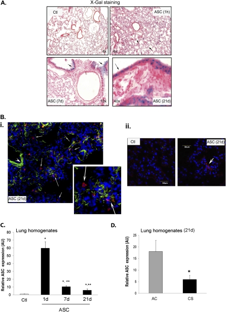
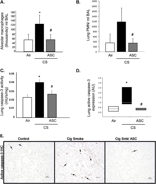
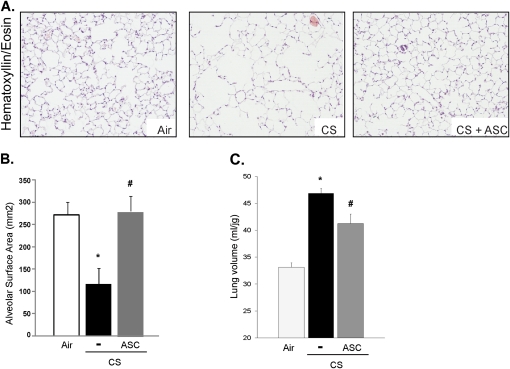
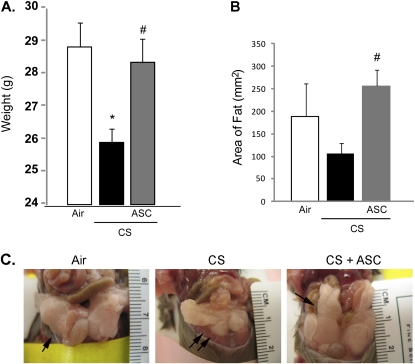
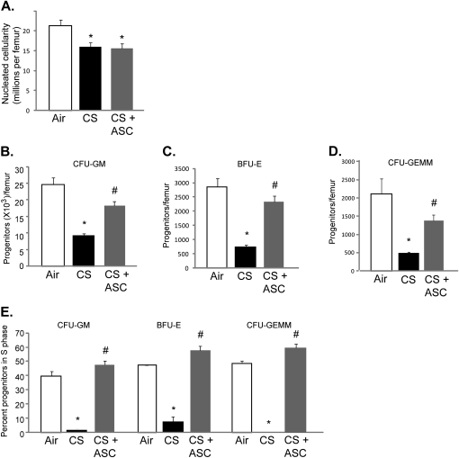
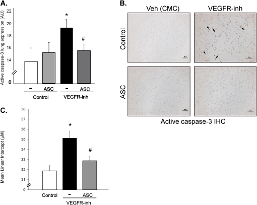
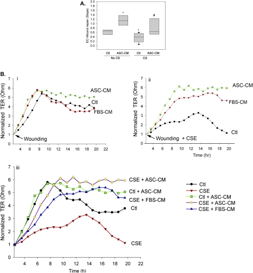
Measurements and main results: Adipose stem cells were detectable in the parenchyma and large airways of lungs up to 21 days after injection. Adipose stem cell treatment was associated with reduced inflammatory infiltration in response to cigarette smoke exposure, and markedly decreased lung cell death and airspace enlargement in both models of emphysema. Remarkably, therapeutic results of adipose stem cells extended beyond lung protection by rescuing the suppressive effects of cigarette smoke on bone marrow hematopoietic progenitor cell function, and by restoring weight loss sustained by mice during cigarette smoke exposure. Pulmonary vascular protective effects of adipose stem cells were recapitulated by application of cell-free conditioned medium, which improved lung endothelial cell repair and recovery in a wound injury repair model and antagonized effects of cigarette smoke in vitro.

Conclusions: These results suggest a useful therapeutic effect of adipose stem cells on both lung and systemic injury induced by cigarette smoke, and implicate a lung vascular protective function of adipose stem cell derived paracrine factors.


Figures

Figure 1.
Figure 1.
Lung homing of adult adipose stem cells (ASC) delivered by intravenous administration to mice. (A) Localization of B-galactosidase–expressing murine ASC (blue) on lung sections imaged at the indicated magnification after fixation and staining with X-Gal and hematoxylin. Lungs of Apo E mice were harvested at the indicated time (1 h, 7 and 21 d) after 5 × 10⁵ ASC or control vehicle (Ctl) administration. Note (arrows) the presence of ASC in the lung parenchyma (1 h) and among the bronchial epithelial layer (7 and 21 d). (B) Epifluorescence (i; inset) and confocal fluorescence (ii) micrographs of frozen-sectioned lungs from DBA/2J mice harvested 21 days after a single injection with DiI-labeled ASC (3 × 10⁵) or vehicle (controls) and stained with DAPI (blue). Note perinuclear red fluorescence (arrows) in the ASC-injected mice, and green autofluorescence of elastin structures in the lung (arrowhead in i). (C) Abundance of DiI-labeled murine ASC detected by flow cytometry of whole-lung homogenates obtained by digestion and disintegration. Lungs were harvested 1, 7, and 21 days after ASC administration (3 × 10⁵) or vehicle in DBA/2J mice previously exposed to cigarette smoking for 2 weeks. All ASC groups *P < 0.05 versus vehicle control; **P < 0.05 versus day 1; n = 4–5; analysis of variance. (D) Abundance of cell events consistent with DiI-labeled murine ASC detected by flow cytometry of lung homogenates harvested 21 days after a single ASC administration (3 × 10⁵) or vehicle in DBA/2J mice previously exposed to cigarette smoking (CS) or air control (AC) for 2 weeks. Mean + SEM; *P < 0.05 versus AC; n = 4–5.
Figure 2.
Figure 2.
Effect of adipose stem cells (ASC) treatment on cigarette smoking (CS)–induced inflammation and caspase activation. Abundance of inflammatory cells alveolar macrophages (A) and polymorphonuclear cells (B) in the bronchoalveolar lavage (BAL) fluid collected from DBA/2J mice exposed to CS or ambient air (Air) for 4 months (n = 8–12 per group) and treated with ASCs (3 × 10⁵ cells infused intravenously every other week, during mo 3 and 4 of CS exposure). *P < 0.05 versus control; #P < 0.05 versus CS; analysis of variance (ANOVA). Lung cell death was quantified in the same experiment by caspase-3 activity measured with a fluorimetric enzymatic kinetic assay and normalized by protein concentration in lung homogenates (C; mean + SEM; *P < 0.05 vs. vehicle control; ANOVA) and by abundance of active caspase-3-expressing cells in lung parenchyma measured (D; median box plot; arbitrary units [AU]; *P < 0.05 vs. air control; #P < 0.05 vs. CS; ANOVA) by automated image analysis of lung sections immunostained with a specific antibody (E). Note active caspase-3 expressing cells in the alveolar tissue (arrows).
Figure 3.
Figure 3.
Effect of adipose stem cells (ASC) treatment on cigarette smoking (CS)–induced airspace enlargement. (A) Alveolar airspaces stained with hematoxylin and eosin on fixed lung sections from mice exposed to CS or ambient air for 4 months. DBA/2J mice were treated with ASC (3 × 10⁵ cells per injection, injected intravenously every other week), during months 3 and 4 of CS exposure. Note the increased airspaces in the CS-exposed mice and the smaller airspaces in the CS-exposed mice treated with ASC. (B) Alveolar surface area calculated by standardized morphometry of alveolar spaces on coded slides (mean + SEM; *P < 0.05 vs. air control; #P < 0.05 vs. CS; analysis of variance). (C) Lung volumes measured in anesthetized and intubated DBA/2J mice (n = 5–10) at 4 months after CS exposure (mean + SEM; *P < 0.05 vs. air control; #P < 0.05 vs. CS; analysis of variance).
Figure 4.
Figure 4.
Effect of adipose stem cells (ASC) treatment on cigarette smoking (CS)–induced weight loss in mice. (A) Body weight of DBA/2J mice after 4 months of air or CS exposure. A third group was treated with ASC (3 × 10⁵ cells per injection, injected intravenously every other week), during months 3 and 4 of CS exposure (mean +SEM; n = 10–12; *P < 0.05 vs. air control; #P < 0.05 vs. CS; analysis of variance). (B) Abundance of abdominal fat (mean + SEM; n = 3–6; #P < 0.05 vs. CS; analysis of variance), measured at 4 months. (C). Note the marked decrease in the amount of abdominal fat in the CS-exposed mice (double arrows), compared with control mice and with ASC-treated CS-exposed mice (arrows).
Figure 5.
Figure 5.
Effect of adipose stem cells (ASC) treatment on cigarette smoking (CS)–induced bone marrow (BM) dysfunction in mice. Absolute numbers of nucleated cells (A) and hematopoietic progenitors colony forming unit- granulocyte, monocyte (CFU-GM) (B), burst-forming unit-erythroid (BFU-E) (C), colony forming unit-granulocyte, erythrocyte, monocyte, and megakaryocyte, CFU-GEMM (D), and cycling status (= percent cells in S-phase) of these progenitors (E) in DBA/2J mice after 4 months of air or CS exposure, with a third group treated with ASC (3 × 10⁵ cells per injection, injected intravenously every other week), during months 3 and 4 of CS exposure (mean + SEM; n = 4–6; *P < 0.05 vs. air control; #P < 0.005 vs. CS; analysis of variance).
Figure 6.
Figure 6.
Adipose stem cells (ASC) efficacy in an apoptosis–dependent alveolar enlargement model. (A, B) Lung cell death was quantified by abundance of active caspase-3 expressing cells in lung parenchyma (at 4 wk) in animals (Nod-SCID NS2 mice) who received a single dose of vascular endothelial growth factor receptor (VEGFR) inhibitor (SU5416, 20 mg/kg; sq) or its vehicle control (carboxymethylcellulose), and who were treated with human adult ASC (3 × 10⁵, intravenous injection) on Day 3 after VEGFR inhibition; (A; mean arbitrary units (AU) + SEM; *P < 0.05 vs. vehicle [control]; #P < 0.05 vs. ASC-untreated [-] animals who received the VEGFR-inhibitor; analysis of variance). Quantification was achieved by image analysis of lung sections immunostained with a specific active caspase-3 antibody (B; brown; arrows). (C) Mean linear intercepts calculated by standardized morphometry of alveolar spaces stained with hematoxylin and eosin from mice exposed to carboxymethylcellulose vehicle or VEGFR-inhibitor for 24 weeks and treated with ASC as above (mean + SEM; *P < 0.05 vs. vehicle control; #P < 0.05 vs. VEGFR-inhibited mice; analysis of variance).
Figure 7.
Figure 7.
Paracrine effects of adipose stem cells (ASC) on lung endothelia injury in vitro. (AC) Wound injury repair measured by the recovery of transcellular electrical resistance (TER) across a confluent monolayer of primary human lung microvascular endothelial cells grown on gold microelectrodes using the Electric Cell-Substrate Impedance Sensing system. A linear electrical injury was applied at time 2.4 hours (B, C, arrow) and the slope of TER recovery to plateau was compared for cells maintained in their regular growth medium, or in medium supplemented with conditioned medium from ASC cells (ASC-CM; 50%), in the absence and presence of cigarette smoking (CS) extract (4%). (A) Box plot with medians; n = 4 independent experiments; P < 0.01 two-way analysis of variance for the effect of CS and ASC-CM; *P < 0.005 versus untreated wounded control cells; #P < 0.005 versus untreated wounded CS-exposed cells. (B) Kinetics of normalized TER (to the TER at time of wound application) (mean; n = 3–4 independent experiments) in unexposed cells (i) or in cells exposed to CS (ii) wounded at time 2.4 hours (arrow), which were either untreated, grown in their control medium (Ctl; black line), or treated with ASC-CM (green line) or with control serum-containing media (fetal bovine serum [FBS]-CM, 20%; red line). Note the effect of CS extract on both the slope and the attained plateau levels of TER recovery in wounded lung endothelial cells and the protective effects of both ASC-CM and serum on the slope of TER recovery, with ASC-CM-specific effects on the plateau TER, evident in panel (iii), which combines conditions shown in panels i and ii.

Similar articles

Cited by

References

    1. Kasahara Y, Tuder RM, Taraseviciene-Stewart L, Le Cras TD, Abman S, Hirth PK, Waltenberger J, Voelkel NF. Inhibition of VEGF receptors causes lung cell apoptosis and emphysema. J Clin Invest 2000;106:1311–1319. - PMC - PubMed
    1. Diab KJ, Adamowicz JJ, Kamocki K, Rush NI, Garrison J, Gu Y, Schweitzer KS, Skobeleva A, Rajashekhar G, Hubbard WC, et al. Stimulation of sphingosine 1 phosphate signaling as an alveolar cell survival strategy in emphysema. Am J Respir Crit Care Med 2010;181:344–352. - PMC - PubMed
    1. Petrache I, Natarajan V, Zhen L, Medler TR, Richter AT, Cho C, Hubbard WC, Berdyshev EV, Tuder RM. Ceramide upregulation causes pulmonary cell apoptosis and emphysema-like disease in mice. Nat Med 2005;11:491–498. - PMC - PubMed
    1. Giordano RJ, Lahdenranta J, Zhen L, Chukwueke U, Petrache I, Langley RR, Fidler IJ, Pasqualini R, Tuder RM, Arap W. Targeted induction of lung endothelial cell apoptosis causes emphysema-like changes in the mouse. J Biol Chem 2008;283:29447–29460. - PMC - PubMed
    1. Cai L, Johnstone BH, Cook TG, Liang Z, Traktuev D, Cornetta K, Ingram DA, Rosen ED, March KL. Suppression of hepatocyte growth factor production impairs the ability of adipose-derived stem cells to promote ischemic tissue revascularization. Stem Cells 2007;25:3234–3243. - PubMed

Publication types

MeSH terms