. 2010 Oct;192(20):5363-77.
doi: 10.1128/JB.00883-10. Epub 2010 Aug 13.

Comparative genome biology of a serogroup B carriage and disease strain supports a polygenic nature of meningococcal virulence

Affiliations

Comparative genome biology of a serogroup B carriage and disease strain supports a polygenic nature of meningococcal virulence

Biju Joseph et al. J Bacteriol. 2010 Oct.

Abstract

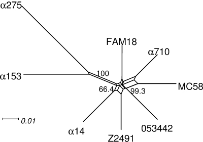
Neisseria meningitidis serogroup B strains are responsible for most meningococcal cases in the industrialized countries, and strains belonging to the clonal complex ST-41/44 are among the most prevalent serogroup B strains in carriage and disease. Here, we report the first genome and transcriptome comparison of a serogroup B carriage strain from the clonal complex ST-41/44 to the serogroup B disease strain MC58 from the clonal complex ST-32. Both genomes are highly colinear, with only three major genome rearrangements that are associated with the integration of mobile genetic elements. They further differ in about 10% of their gene content, with the highest variability in gene presence as well as gene sequence found for proteins involved in host cell interactions, including Opc, NadA, TonB-dependent receptors, RTX toxin, and two-partner secretion system proteins. Whereas housekeeping genes coding for metabolic functions were highly conserved, there were considerable differences in their expression pattern upon adhesion to human nasopharyngeal cells between both strains, including differences in energy metabolism and stress response. In line with these genomic and transcriptomic differences, both strains also showed marked differences in their in vitro infectivity and in serum resistance. Taken together, these data support the concept of a polygenic nature of meningococcal virulence comprising differences in the repertoire of adhesins as well as in the regulation of metabolic genes and suggest a prominent role for immune selection and genetic drift in shaping the meningococcal genome.


Figures

FIG. 1.
FIG. 1.
Neighbor net based on the distribution of 2,295 COGs in eight meningococcal genomes. The numbers at each split give the percent bootstrap support in 1.000 replicate. The data sets suggest that the α710 genome is closer to the MC58 genome than to the other meningococcal genomes from non-serogroup B strains.
FIG. 2.
FIG. 2.
Whole-genome and transcriptome comparisons of the two serogroup B meningococcal strains. The linearized α710 and MC58 genomes are shown in the middle-upper and middle-lower panels as gray bars, and regions syntenic in both genomes are connected via red and inverted regions via blue lines, respectively. Above and below the aligned genomes, the Karlin signature differences are given to detect regions of atypical compositional bias in both genomes, such as IHTs or the ribosomal operons (R). In addition, at the upper and lower margins the log2-fold changes in gene expression are given for both genomes, comparing the transcriptomes of cells in mid-log growth in RPMI 1640 medium and upon adhesion to human FaDu nasopharyngeal cell using a MC58-based microarray. In the α710 genome, black vertical lines on the forward and reverse strand indicate the positions of the five gaps (I to V). Pink boxes termed IT1, IT2, and IT3 designate inversions coupled to translocations as described in the text, a chromosomal region duplicated in the genome of MC58 (D), and the position of a P2-like prophage. The TPS system-encoding locus missing in the α710 genome is indicated by a pink box in the MC58 genome panel, and the differing positions of the Nf1 prophages are indicated by a red box in both genomes. Loci that have an atypical nucleotide composition and that contain a number of transcriptionally silent gene cassettes (discussed in more detail in the text) are given as green boxes.
FIG. 3.
FIG. 3.
Distribution of strain-specific and core genes in pairwise genome comparisons among the different functional categories and cellular compartments. (A) Histogram depicting the COG functional category profile for strain-specific and shared genes. Genes present in both genomes differ in their functional category profile from strain-specific genes (P < 0.01, χ2 test). The latter are enriched for genes that do not belong to any functional category (COG X), that code for proteins involved in cell motility (COG N), or that code for proteins involved in replication, recombination, and repair (COG L) (P = 0.055) (*, P < 0.05; **, P < 0.01; Fisher's exact test with BH multiple testing correction). Abbreviations: C, energy production and conversion; D, cell cycle control, mitosis, and meiosis; E, amino acid transport and metabolism; F, nucleotide transport and metabolism; G, carbohydrate transport and metabolism; H, coenzyme transport and metabolism; I, lipid transport and metabolism; J, translation; K, transcription; L, replication, recombination, and repair; M, cell wall/membrane biogenesis; N, cell motility; O, posttranslational modification, protein turnover, and chaperones; P, inorganic ion transport and metabolism; Q, secondary metabolite biosynthesis, transport, and catabolism; R, general function prediction only; S, function unknown; T, signal transduction mechanisms; U, intracellular trafficking and secretion; V, defense mechanisms; X, not in COGs. (B) Histogram showing the different distributions of strain-specific and shared genes among the different subcellular compartments (P < 0.01, χ2 test) as predicted by PSORTb. Common genes are enriched for genes coding for cytoplasmic or inner membrane proteins, respectively (*, P < 0.05; **, P < 0.01; Fisher's exact test with BH multiple testing correction). Abbreviations: CP, cytoplasmic; CM, cytoplasmic membrane; PP, periplasmic; OM, outer membrane; EC, extracellular; UK, unknown.
FIG. 4.
FIG. 4.
Comparison of the sequence variability between orthologs as expressed by their BSRs among the different COG functional categories and subcellular compartments. (A) Box-and-whiskers plot of the BSRs grouped by COG categories. In addition to significant differences among the different COG categories (P < 0.01, Kruskal-Wallis test), the levels of BSRs of orthologs belonging to the COG functional categories J and K are significantly higher and those belonging to category X are significantly lower than the overall BSR median, which is indicated by a black horizontal line (*, P < 0.05; two-sided Wilcoxon test with BH multiple testing correction). The 95% quantile (dashed horizontal line) is given to separate the 5% most variable orthologous pairs. For the abbreviations of the COG functional categories, see the legend to Fig. 3A. (B) Box-and-whiskers plot of the BSRs grouped by subcellular localization as predicted by PSORTb. Again, there are significant BSR differences between the subcellular compartments (P = 0.016, Kruskal-Wallis test) and compared to the overall BSR median (black horizontal line). Outer membrane proteins have a significantly lower degree of sequence conservation (*, P < 0.05; two-sided Wilcoxon test with BH multiple testing correction). For the abbreviations of the different subcellular compartments, see the legend to Fig. 3B. (C) Histogram comparing the COG functional distribution of the 5% orthologs with the highest BSRs and the remaining 95% of more-conserved orthologs from panel A. Whereas the more-conserved orthologs belong more often to the COG functional categories E and J, the 5% most-variable orthologs are more enriched for the COG functional categories U and X than the former (*, P < 0.05; **, P < 0.01; Fisher's exact test with BH multiple testing correction). (D) Histogram showing the distribution of the 5% of genes with the highest BSRs among the different subcellular compartments as predicted by PSORTb from panel B. Whereas the more-conserved orthologs are located significantly more frequently in the cytoplasm than the 5% most-variable orthologs, the latter are more often located at the outer membrane (P = 0.052) or have no subcellular localization prediction (*, P < 0.05; **, P < 0.01; Fisher's exact test with BH multiple testing correction). For the abbreviations, refer to the legend to Fig. 3.
FIG. 5.
FIG. 5.
Functional classification of genes differentially expressed upon adhesion to human FaDu nasopharyngeal cell lines in the two meningococcal serogroup B strains MC58 and α710. Core genes regulated in both strains are depicted as black bars, core genes regulated only in MC58 as dark gray, and those regulated only in α710 as light gray bars, respectively; regulated MC58-specific genes are depicted as white bars. (A) Distribution of core and MC58-specific genes among the five different COG functional classes. The regulated core genes are not distributed equally over the five functional classes (P < 0.01, χ2 test) but are enriched for metabolic genes in the respective three datasets (see Table S4 in the supplemental material). Likewise, the distribution of core genes that are regulated in only one strain also differs significantly between both strains (P < 10−15, χ2 test) and also from the core genes regulated in both strains (P < 10−9, χ2 test). In contrast, the regulated MC58-specific genes are evenly distributed over all functional classes (P = 0.77, χ2 test). (B) Distribution among the different COG functional categories separated into up- and downregulated genes. For each of the four datasets, the percentage of regulated genes adds up to 100% separately. Fifty-eight percent of the downregulated MC58-specific genes fall into COG category X, which therefore falls off the scale to the right side of the histogram. In all four datasets the downregulated genes are enriched for genes not in COGs (COG X), whereas the core genes that are upregulated in only one strain are enriched for genes coding for proteins involved in translation, ribosomal structure, and biogenesis (COG J) (P < 0.05; Fisher's exact test with BH multiple testing correction). For clarity, significant differences have not been depicted in the diagram. For the abbreviations, refer to the legend to Fig. 3.

Similar articles

Cited by

References

    1. Altschul, S. F., W. Gish, W. Miller, E. W. Myers, and D. J. Lipman. 1990. Basic local alignment search tool. J. Mol. Biol. 215:403-410. - PubMed
    1. Arber, W. 2000. Genetic variation: molecular mechanisms and impact on microbial evolution. FEMS Microbiol. Rev. 24:1-7. - PubMed
    1. Bentley, S. D., G. S. Vernikos, L. A. Snyder, C. Churcher, C. Arrowsmith, T. Chillingworth, A. Cronin, P. H. Davis, N. E. Holroyd, K. Jagels, M. Maddison, S. Moule, E. Rabbinowitsch, S. Sharp, L. Unwin, S. Whitehead, M. A. Quail, M. Achtman, B. Barrell, N. J. Saunders, and J. Parkhill. 2007. Meningococcal genetic variation mechanisms viewed through comparative analysis of serogroup C strain FAM18. PLoS Genet. 3:e23. - PMC - PubMed
    1. Bourdoulous, S., and X. Nassif. 2006. Mechanisms of attachment and invasion, p. 257-272. In M. Frosch and M. C. Maiden (ed.), Handbook of meningococcal disease. Wiley-VCH, Weinheim, Germany.
    1. Buckee, C. O., K. A. Jolley, M. Recker, B. Penman, P. Kriz, S. Gupta, and M. C. Maiden. 2008. Role of selection in the emergence of lineages and the evolution of virulence in Neisseria meningitidis. Proc. Natl. Acad. Sci. U. S. A. 105:15082-15087. - PMC - PubMed

Publication types

MeSH terms

Substances