. 2010 Oct;192(20):5390-401.
doi: 10.1128/JB.00711-10. Epub 2010 Aug 13.

AmrZ beta-sheet residues are essential for DNA binding and transcriptional control of Pseudomonas aeruginosa virulence genes

Affiliations

AmrZ beta-sheet residues are essential for DNA binding and transcriptional control of Pseudomonas aeruginosa virulence genes

Elizabeth A Waligora et al. J Bacteriol. 2010 Oct.

Abstract

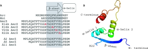
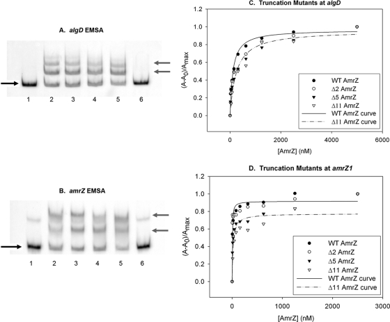
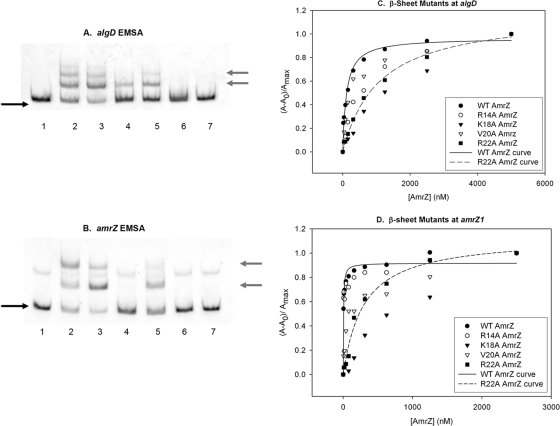
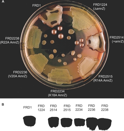
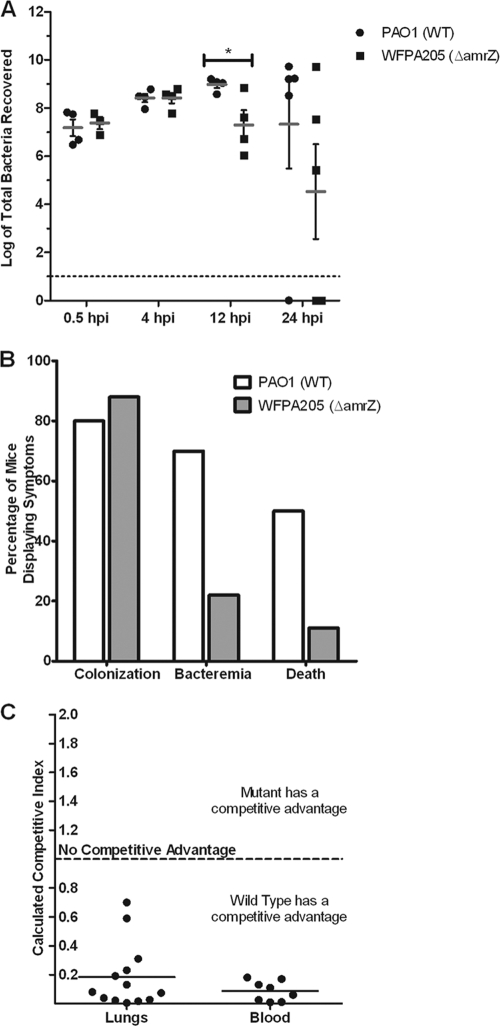
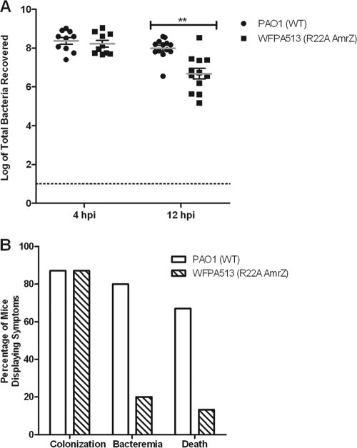
AmrZ is a putative ribbon-helix-helix (RHH) transcriptional regulator. RHH proteins utilize residues within the β-sheet for DNA binding, while the α-helices promote oligomerization. AmrZ is of interest due to its dual roles as a transcriptional activator and as a repressor, regulating genes encoding virulence factors associated with both chronic and acute Pseudomonas aeruginosa infection. In this study, cross-linking revealed that AmrZ forms oligomers in solution but that the amino terminus, containing an unordered region and a β-sheet, were not required for oligomerization. The first 12 unordered residues (extended amino terminus) contributed minimally to DNA binding. Mutagenesis of the AmrZ β-sheet demonstrated that residues 18, 20, and 22 were essential for DNA binding at both activation and repressor sites, suggesting that AmrZ utilizes a similar mechanism for binding to these sites. Mice infected with amrZ mutants exhibited reduced bacterial burden, morbidity, and mortality. Direct in vivo competition assays showed a 5-fold competitive advantage for the wild type over an isogenic amrZ mutant. Finally, the reduced infection phenotype of the amrZ-null strain was similar to that of a strain expressing a DNA-binding-deficient AmrZ variant, indicating that DNA binding and transcriptional regulation by AmrZ is responsible for the in vivo virulence defect. These recent infection data, along with previously identified AmrZ-regulated virulence factors, suggest the necessity of AmrZ transcriptional regulation for optimal virulence during acute infection.


Figures

FIG. 1.
FIG. 1.
Alignment and predicted secondary structure of the putative ribbon-helix-helix transcriptional regulator AmrZ. (A) An amino acid alignment of the Arc-like DNA-binding domains of Arc (residues 1 to 18), Mnt (residues 1 to 15), and AmrZ (residues 1 to 27) reveals conserved residues in the DNA-binding β-sheet as well as the presence of the extended amino terminus. Residues in gray indicate the DNA-binding β-sheet. Residues to the left are part of the extended amino acid. Residues within the DNA-binding β-sheet that were targeted for site-specific mutagenesis are shown in red. (B) Predicted three-dimensional (3D) structure of AmrZ residues 13 to 80 provided by the secondary-structure prediction program pyMol. The major structural components (N and C termini, β-sheet, and α-helices) are indicated. The location of arginine-22 (R22) is also indicated, given the frequent references to the R22A mutant.
FIG. 2.
FIG. 2.
Purified AmrZ forms oligomers in solution, and oligomerization does not require the first 26 residues. (A and B) Purification and verification of the wild type and the extended amino truncation proteins Δ2AmrZ, Δ5AmrZ, and Δ11AmrZ by SDS-PAGE and visualization by GelCode staining (A) and Western blotting (B). Lane 2, wild-type AmrZ; lane 3, Δ2AmrZ; lane 4, Δ5AmrZ; lane 5, Δ11AmrZ. (C and D) Purification and verification in parallel of the wild type and the R14A AmrZ (lane 3), K18A AmrZ (lane 4), V20A AmrZ (lane 5), and R22A AmrZ (lane 6) β-sheet mutants by SDS-PAGE and visualization by GelCode staining (C) and Western blotting (D). (E) Samples were cross-linked by incubation with glutaraldehyde to form a stable complex that would withstand separation by SDS-PAGE. Lanes 1 to 6 are the β-sheet mutants. AmrZ in lane 1 was not incubated with glutaraldehyde to indicate the size of a wild-type AmrZ monomer. Lanes 2 to 6 are cross-linked and contain wild-type AmrZ (lane 2), R14A AmrZ (lane 3), K18A AmrZ (lane 4), V20A AmrZ (lane 5), and R22A AmrZ (lane 6). Lanes 8 to 15 represent the truncation proteins with alternating untreated monomeric and glutaraldehyde-treated samples. Lanes 8 and 9, wild-type AmrZ; lanes 10 and 11, Δ2AmrZ; lanes 12 and 13, Δ5AmrZ; lanes 14 and 15, Δ11AmrZ. All lanes contain 40 μmol of protein and were separated by SDS-PAGE on a 12% polyacrylamide gel, and complexes were visualized by GelCode staining.
FIG. 3.
FIG. 3.
The AmrZ extended amino terminus is not required for DNA-binding activity. (A and B) Protein activity was analyzed with 5′-FAM-labeled PCR-amplified targets of the algD (A) or amrZ (B) promoter region. Lanes 1 and 6 have no protein. lane 2, wild-type (WT) AmrZ; lane 3, Δ2AmrZ; lane 4, Δ5AmrZ; lane 5, Δ11AmrZ (at 125 nM each). The black arrows on the left indicate free unbound DNA, while the gray arrows indicate the migration of DNA-protein complexes. The fluorescence anisotropy data (see Materials and Methods) were assembled into a table, separated for each target site (Table 1). (C and D) A representative experiment is illustrated for the activator site algD (C) and the high-affinity repressor site amrZ1 (D).
FIG. 4.
FIG. 4.
Mutation of specific residues causes a loss of AmrZ-mediated DNA-binding activity. (A and B) Protein activity was analyzed with 5′-FAM-labeled PCR-amplified targets of the algD activator (A) or amrZ (B) promoter region. Lanes 1 and 7 have no protein. Lane 2, wild-type AmrZ; lane 3, R14A AmrZ; lane 4, K18A AmrZ; lane 5, V20A AmrZ; lane 6, R22A AmrZ (at 125 nM each). The black arrows on the left indicate free unbound DNA, while the gray arrows indicate the migration of DNA-protein complexes. The fluorescence anisotropy data (see Materials and Methods) were assembled into a table, separated for each target site (Table 1). (C and D) A representative experiment is illustrated for the activator site algD (C) and the high-affinity repressor site amrZ1 (D).
FIG. 5.
FIG. 5.
Residues Lys18, Val20, and Arg22 are required for alginate production. (A) The wild-type amrZ strain (FRD1), an amrZ-null strain (FRD1224), and FRD1224 complemented with wild-type amrZ (FRD2514), amrZ28 (AmrZ R14A) (FRD2515), amrZ17 (AmrZ K18A) (FRD2234), amrZ18 (AmrZ V20A) (FRD2236), or amrZ19 (AmrZ R22A) (FRD2238) was plated onto LANS and incubated at 37°C for 24 h to visualize the alginate phenotype. (B) Western blot of strains expressing the wild type or the null or alanine substitution mutants of AmrZ. Strain names are identical to those listed in A.
FIG. 6.
FIG. 6.
amrZ mutants exhibit an in vivo virulence defect. (A) Wild-type strain PAO1 and strain WFPA205 (amrZ::tet) were used to intranasally inoculate 6-week-old C57BL/6 mice with 1 × 10⁸ bacteria. At the indicated time points (hpi), lungs were aseptically harvested, and the bacteria were enumerated. Each symbol indicates an individual mouse (n = 5 for each group per time point). Gray bars indicate averages along with standard deviations (*, P = 0.036). (B) Mice (n = 15) were assessed for bacterial colonization (>104 bacteria recovered from the lungs), bacteremia (bacteria recovered from the blood), or death (requiring euthanasia due to severe disease symptoms) at 12 hpi. (C) Strains PAO1 and WFPA205 were coinoculated at a 1:1 ratio (total of 108 bacteria). At 24 hpi, lungs and blood were harvested, and bacterial counts were determined. The competitive index from the lungs is plotted for each mouse (n = 13) and for those that showed evidence of bacteremia (n = 8).
FIG. 7.
FIG. 7.
AmrZ DNA-binding activity is required for optimum virulence. (A) A total of 108 PAO1 or WFPA513 (R22A AmrZ) bacteria were used to intranasally inoculate 6-week-old C57BL/6 mice. At 4 and 12 hpi, lungs were aseptically harvested, and the bacteria were enumerated. Each symbol indicates an individual mouse (n = 15 for each group per time point). Gray bars indicate averages along with standard deviations (**, P = 0.003). (B) Mice (n = 15) were assessed for bacterial colonization (>104 bacteria recovered from the lungs), bacteremia (bacteria recovered from the blood), or death (requiring euthanasia due to severe disease symptoms) at 12 hpi.

References

    1. Barillà, D., E. Carmelo, and F. Hayes. 2007. The tail of the ParG DNA segregation protein remodels ParF polymers and enhances ATP hydrolysis via an arginine finger-like motif. Proc. Natl. Acad. Sci. U. S. A. 104:1811-1816. - PMC - PubMed
    1. Baynham, P. J., A. L. Brown, L. L. Hall, and D. J. Wozniak. 1999. Pseudomonas aeruginosa AlgZ, a ribbon-helix-helix DNA-binding protein, is essential for alginate synthesis and algD transcriptional activation. Mol. Microbiol. 33:1069-1080. - PubMed
    1. Baynham, P. J., D. M. Ramsey, B. V. Gvozdyev, E. M. Cordonnier, and D. J. Wozniak. 2006. The Pseudomonas aeruginosa ribbon-helix-helix DNA-binding protein AlgZ (AmrZ) controls twitching motility and biogenesis of type IV pili. J. Bacteriol. 188:132-140. - PMC - PubMed
    1. Baynham, P. J., and D. J. Wozniak. 1996. Identification and characterization of AlgZ, an AlgT-dependent DNA-binding protein required for Pseudomonas aeruginosa algD transcription. Mol. Microbiol. 22:97-108. - PubMed
    1. Benanti, E. L., and P. T. Chivers. 2007. The N-terminal arm of the Helicobacter pylori Ni2+-dependent transcription factor NikR is required for specific DNA binding. J. Biol. Chem. 282:20365-20375. - PubMed

Publication types

LinkOut - more resources