

Gray platelet syndrome: natural history of a large patient cohort and locus assignment to chromosome 3p
- PMID: 20709904
- PMCID: PMC3012593
- DOI: 10.1182/blood-2010-05-286534
Gray platelet syndrome: natural history of a large patient cohort and locus assignment to chromosome 3p
Abstract
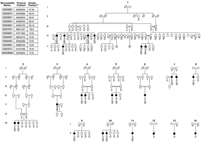
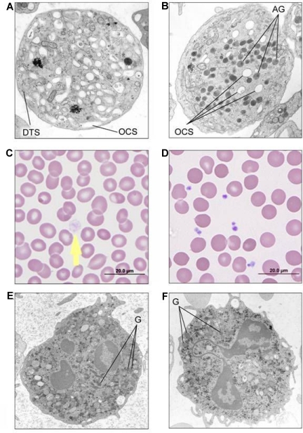
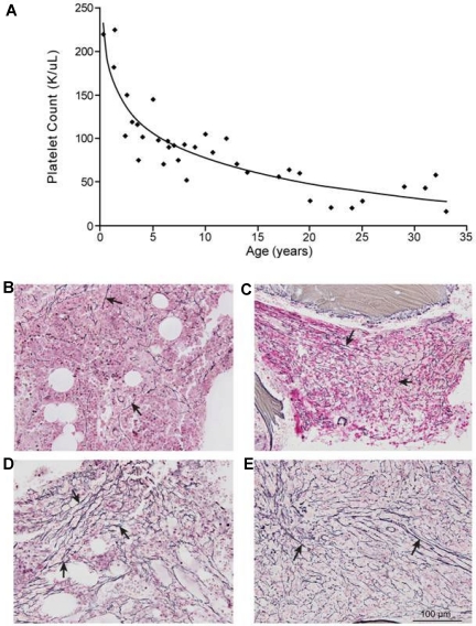
Gray platelet syndrome (GPS) is an inherited bleeding disorder characterized by macrothrombocytopenia and absence of platelet α-granules resulting in typical gray platelets on peripheral smears. GPS is associated with a bleeding tendency, myelofibrosis, and splenomegaly. Reports on GPS are limited to case presentations. The causative gene and underlying pathophysiology are largely unknown. We present the results of molecular genetic analysis of 116 individuals including 25 GPS patients from 14 independent families as well as novel clinical data on the natural history of the disease. The mode of inheritance was autosomal recessive (AR) in 11 and indeterminate in 3 families. Using genome-wide linkage analysis, we mapped the AR-GPS gene to a 9.4-Mb interval on 3p21.1-3p22.1, containing 197 protein-coding genes. Sequencing of 1423 (69%) of the 2075 exons in the interval did not identify the GPS gene. Long-term follow-up data demonstrated the progressive nature of the thrombocytopenia and myelofibrosis of GPS resulting in fatal hemorrhages in some patients. We identified high serum vitamin B(12) as a consistent, novel finding in GPS. Chromosome 3p21.1-3p22.1 has not been previously linked to a platelet disorder; identification of the GPS gene will likely lead to the discovery of novel components of platelet organelle biogenesis. This study is registered at www.clinicaltrials.gov as NCT00069680 and NCT00369421.
Figures
Comment in
-
Filling a void in Gray Platelets.Blood. 2010 Dec 2;116(23):4738-40. doi: 10.1182/blood-2010-09-304378. Blood. 2010. PMID: 21127182 No abstract available.
References
-
- Raccuglia G. Gray platelet syndrome. A variety of qualitative platelet disorder. Am J Med. 1971;51(6):818–828. - PubMed
-
- Nurden AT, Nurden P. The gray platelet syndrome: clinical spectrum of the disease. Blood Rev. 2007;21(1):21–36. - PubMed
-
- Levy-Toledano S, Caen JP, Breton-Gorius J, et al. Gray platelet syndrome: alpha-granule deficiency. Its influence on platelet function. J Lab Clin Med. 1981;98(6):831–848. - PubMed
-
- Jantunen E, Hanninen A, Naukkarinen A, Vornanen M, Lahtinen R. Gray platelet syndrome with splenomegaly and signs of extramedullary hematopoiesis: a case report with review of the literature. Am J Hematol. 1994;46(3):218–224. - PubMed
Publication types
MeSH terms
Substances
Associated data
Grants and funding
LinkOut - more resources
Full Text Sources
Medical
Molecular Biology Databases
Research Materials
Miscellaneous
