. 2010 Dec;24(12):4767-81.
doi: 10.1096/fj.10-154757. Epub 2010 Aug 13.

The melanoma-associated transmembrane glycoprotein Gpnmb controls trafficking of cellular debris for degradation and is essential for tissue repair

Affiliations

The melanoma-associated transmembrane glycoprotein Gpnmb controls trafficking of cellular debris for degradation and is essential for tissue repair

Bing Li et al. FASEB J. 2010 Dec.

Abstract

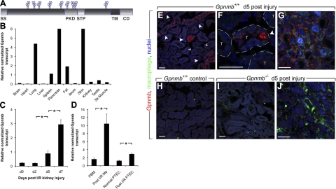
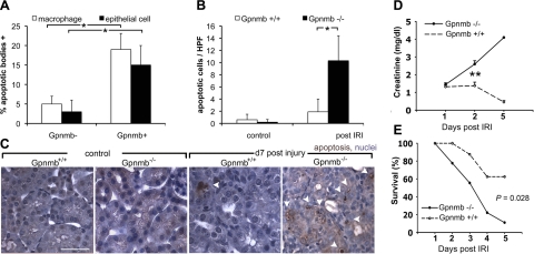
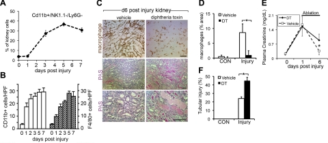
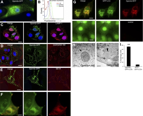
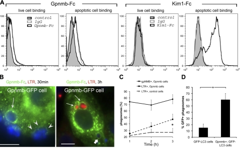
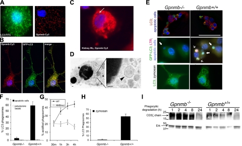
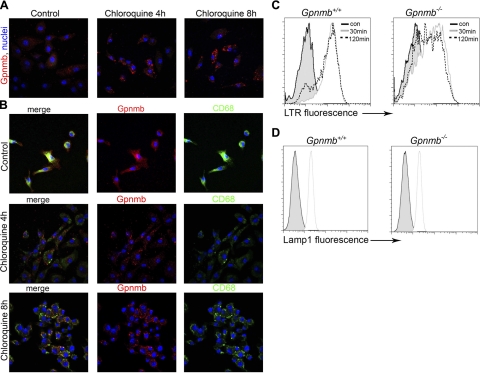
Kidney damage due to injury rarely resolves completely, and there are currently no therapies capable of promoting repair. In addition to understanding mechanisms by which tissues are damaged, illuminating mechanisms of repair and regeneration is also of great importance. Here we show that the melanoma-associated, transmembrane glycoprotein, Gpnmb, is up-regulated 15-fold following ischemic damage in kidney tissue and by more than 10-fold in macrophages and 3-fold in surviving epithelial cells. Gpnmb-expressing macrophages and epithelial cells were found to contain apoptotic bodies at 3 times the rate of nonexpressing cells. Either mutation of Gpnmb or ablation of inflammatory macrophages prevents normal repair of the kidney. Significantly, the kidneys from postischemic Gpnmb mutant mice exhibited a 5-fold increase in apoptotic cellular debris compared to wild-type mice. These mice also experienced an 85% increase in mortality following bilateral ischemic kidney. Finally, we demonstrate that Gpnmb is a phagocytic protein that is necessary for recruitment of the autophagy protein LC3 to the phagosome where these proteins are colocalized and for lysosomal fusion with the phagosome and hence bulk degradation of their content. Therefore, Gpnmb is a novel prorepair gene that is necessary for crosstalk between the macroautophagic degradation pathway and phagocytosis.


Figures

Figure 1.
Figure 1.
Kidney epithelial cells and inflammatory Mφs express Gpnmb in response to ischemic injury. A) Domain model of Gpnmb. B) Representative quantitative PCR (qPCR) of Gpnmb transcripts in normal mouse tissues. C) qPCR for Gpnmb transcript from sham surgery mouse kidney (d 0) and from time points following IRI kidney. D) qPCR for Gpnmb transcript from d 5 post-IRI kidney Mφs compared with peripheral blood monocytes (PBM), and injured proximal tubule epithelial cells (PTEC) compared with normal PTEC. E–J) Confocal images of Gpnmb detected by immunofluorescence in Mφs in normal kidney (H) and d 5 post-IRI mouse kidney outer medulla (E, F) and inner medulla (G). Injured, regenerating epithelial cells express Gpnmb (arrowheads) as well as intratubule epithelial phagocytes. Although inner medulla macrophages coexpress Gpnmb (F), cortical macrophages (E, small arrows) do not. Note Gpnmb+ intracellular vesicles in regenerating epithelial cells (F). T, tubule. In Gpnmb−/− post-IRI kidneys (I, J), outer medulla epithelium (I) and inner medulla Mφs (J) do not express Gpnmb. Scale bars =5 0 μm. *P < 0.05 (n=4/group).
Figure 2.
Figure 2.
Gpnmb is necessary for postischemia reperfusion kidney repair and is required for normal apoptotic cell clearance. A) Proportion of post-IRI d 7 kidney Mφs or epithelial cells in the outer medulla containing apoptotic bodies segregated by expression of Gpnmb. *P < 0.05. B, C) Apoptotic cells seen in the normal and d 7 postunilateral IRI kidneys of Gpnmb−/− mice and controls. Note an abundance of apoptotic bodies (arrowheads) in the kidney outer medulla. D) Plasma creatinine levels in Gpnmb−/− and Gpnmb+/+ mice following bilateral IRI (n=9/group). **P < 0.001. E) Kaplan-Meier survival curves for Gpnmb−/− and Gpnmb+/+ mice following bilateral IRI. P = 0.028
Figure 3.
Figure 3.
Mφ ablation in Cd11b-DTR mice prevents normal kidney repair following IRI. A) Percentage of kidney cells that are CD11b+, NK1.1, Ly6G Mφs, as assessed by flow cytometry. B) CD11b+ and F4/80+ cells in the kidney following IRI, quantified by immunostaining of kidney sections. C) Photomicrographs of F4/80 staining and PAS staining of kidneys at d 6 post-IRI following no Mφ ablation (vehicle) or Mφ ablation with DT from d 3 to 6. D) F4/80 area in control kidneys or d 6 post-IRI kidneys following ablation. E) Effect of Mφ ablation from d 3 to 6 post-IRI on plasma creatinine. Note that following Mφ ablation there is a higher creatinine level on d 6 than in mice with normal Mφ compliment. F) tubule injury score in d 6 post-IRI kidneys following ablation. Scale bars = 100 μm. *P < 0.05.
Figure 4.
Figure 4.
Gpnmb localizes to autophagosomes in epithelial cells and is expressed at low levels at the plasma membrane. A) Gpnmb-GFP is localized to membranes of a network of intracellular vesicles when expressed in kidney epithelial cells in vitro. B) Cell surface expression of Gpnmb, assessed by flow cytometry. Cells were stained either with rabbit IgG (control) or anti-Gpnmb ectodomain-specific antibodies. C) Confocal images of Gpnmb-GFP (green) in cells expressing the transgene and costained with antibodies against LAMP-1 (red). Note that despite extensive networks of both LAMP-1 and Gpnmb compartments, there is no colocalization. D) Confocal images of Gpnmb-GFP-expressing cells stained with LysoTracker Red. Note again there is no colocalization. E) Confocal images of Gpnmb-GFP cells stained with antibodies against the early endosomal marker EEA1 (red). Note there is no colocalization. F) Fluorescence images of Gpnmb-RFP cells stained with the cholesterol marker filipin (false green). Note the extensive colocalization. G) Fluorescence images of Gpnmb-RFP+ GFP-LC3 cells (top panels), and control GFP-LC3 cells (bottom panels). Note extensive colocalization of these 2 proteins and the reorganization of LC3 in Gpnmb-expressing cells. H) Electron micrographs showing the double membrane autophagosome in the cytoplasm of a Gpnmb expressing cell (arrow), and absence of autophgosomes, but the presence of lipid droplets (arrowhead) in control cell. I) Percentage of autophagic cells stably coexpressing Gpnmb-RFP and GFP-LC3 compared with cells expressing GFP-LC3 and control vector RFP. Scale bars = 10 μm (A–G); 100 nm (H).
Figure 5.
Figure 5.
Gpnmb is recruited to the phagocytic cup and directs phagocytic trafficking to a lysosomal degradation pathway A) Binding of Gpnmb-Fc or IgG to live thymocytes or apoptotic thymocytes (left panels) and binding of Kim1-Fc or IgG (right panels). Kim1-Fc specifically binds to apoptotic cells, whereas Gpnmb-Fc does not. B) Gpnmb-GFP-expressing epithelial cells incubated with apoptotic thymocytes for 30 min or 1 h, followed by 2 h incubation. Gpnmb-GFP is recruited to an intact phagosome (arrow) and is also seen at the base of the phagocytic cup (arrowheads) before internalization. Image shows apoptotic bodies within Gpnmb phagosomes at 3 h (arrows). Early phagosomes are not stained with LysoTracker Red (thick arrow) whereas late phagosomes are (thin arrows). C) Time course of Gpnmb association with phagosomes compared with LysoTracker Red association with phagosomes in Gpnmb-GFP cells or control cells. Note that cells not expressing Gpnmb-GFP fail to acidify phagosomes. D) Graph of GFP-LC3 recruitment to phagosomes after 1 h of incubation in GFP-LC3 cells compared with Gpnmb+ GFP-LC3 cells. *P < 0.05 (n=5/group).
Figure 6.
Figure 6.
Endogenous Mφ Gpnmb colocalizes with autophagic proteins and promotes phagosome acidification and degradation. A) Representative confocal images of BMMφs showing endogenously expressed LC3 and Gpnmb localization to intracellular vesicles. B) Representative confocal image of BMMφs transduced with GFP-LC3 and labeled with anti-Gpnmb antibodies, revealing a high degree of colocalization. C) Representative image of kidney Mφs from d 5 post-IRI kidney labeled with antibodies to Gpnmb (red) and colabeled with DAPI. Note the apoptotic body in a Gpnmb phagosome (arrow). D) Electron micrograph of healthy BMMφs showing double-membraned autophagosome adjacent to a single-membrane, apoptotic body phagosome. E) Confocal images of native LC3 immunostaining (red; top panels) or retrovirally transduced GFP-LC3 (green; middle and bottom panels) after 1 h of phagocytosis of apoptotic cells (blue) or zymosan by Gpnmb−/− or Gpnmb+/+ BMMφs. Note that the phagosome frequently contains membrane-associated LC3 in WT Mφs but rarely in Gpnmb−/− Mφs. Cells were coincubated with LysoTracker Red. Note some LC3+ phagosomes in Gpnmb+/+ Mφs show red color, indicative of early lysosome interaction. F) Percentage apoptotic body or polystyrene bead phagosomes in Gpnmb−/− or Gpnmb+/+ Mφs that show ring enhancement due to LC3 in the membrane after 1 h incubation. G) Percentage of zymosan phagosomes in Gpnmb−/− or Gpnmb+/+ Mφs that show ring enhancement due to LC3 in the membrane. H) Percentage apoptotic body phagosomes that label with LysoTracker Red with time in Gpnmb−/− or Gpnmb+/+ Mφs. I) Immunoblot measuring Mφ content of the thymocyte-specific protein CD3 ζ-chain at time points following 1 h of phagocytosis of apoptotic thymocytes. Scale bars = 100 nm (D); 50 μm (E).
Figure 7.
Figure 7.
Characterization of intracellular compartments in Mφs. A) Confocal images of d 7 BMMφs, untreated or treated with chloroquine, then immunolabeled for Gpnmb. B) Split-panel confocal images of CD68 and Gpnmb in BMMφs untreated or treated with chloroquine. C, D) Flow cytometric histogram plots of LysoTracker Red fluorescence (C) and LAMP-1 immunofluorescnce (D) in WT and Gpnmb mutant BMMφs after 30 or 120 min loading. Note a population of LTR dim cells in Gpnmb mutants.

Similar articles

Cited by

References

    1. Duffield J. S., Hong S., Vaidya V. S., Lu Y., Fredman G., Serhan C. N., Bonventre J. V. (2006) Resolvin D series and protectin D1 mitigate acute kidney injury. J. Immunol. 177, 5902–5911 - PubMed
    1. Ichimura T., Asseldonk E. J., Humphreys B. D., Gunaratnam L., Duffield J. S., Bonventre J. V. (2008) Kidney injury molecule-1 is a phosphatidylserine receptor that confers a phagocytic phenotype on epithelial cells. J. Clin. Invest. 118, 1657–1668 - PMC - PubMed
    1. Castano A. P., Lin S. L., Surowy T., Nowlin B. T., Turlapati S. A., Patel T., Singh A., Li S., Lupher M. L., Jr., Duffield J. S. (2009) Serum amyloid P inhibits fibrosis through Fc gamma R-dependent monocyte-macrophage regulation in vivo. Sci. Transl. Med. 1, 5ra13 - PMC - PubMed
    1. Arnold L., Henry A., Poron F., Baba-Amer Y., van Rooijen N., Plonquet A., Gherardi R. K., Chazaud B. (2007) Inflammatory monocytes recruited after skeletal muscle injury switch into antiinflammatory macrophages to support myogenesis. J. Exp. Med. 204, 1057–1069 - PMC - PubMed
    1. Duffield J. S., Forbes S. J., Constandinou C. M., Clay S., Partolina M., Vuthoori S., Wu S., Lang R., Iredale J. P. (2005) Selective depletion of macrophages reveals distinct, opposing roles during liver injury and repair. J. Clin. Invest. 115, 56–65 - PMC - PubMed

Publication types

MeSH terms

LinkOut - more resources