. 2010 Aug 15;70(16):6412-9.
doi: 10.1158/0008-5472.CAN-10-0052.

Type 1 insulin-like growth factor receptor translocates to the nucleus of human tumor cells

Affiliations

Type 1 insulin-like growth factor receptor translocates to the nucleus of human tumor cells

Tamara Aleksic et al. Cancer Res. .

Abstract

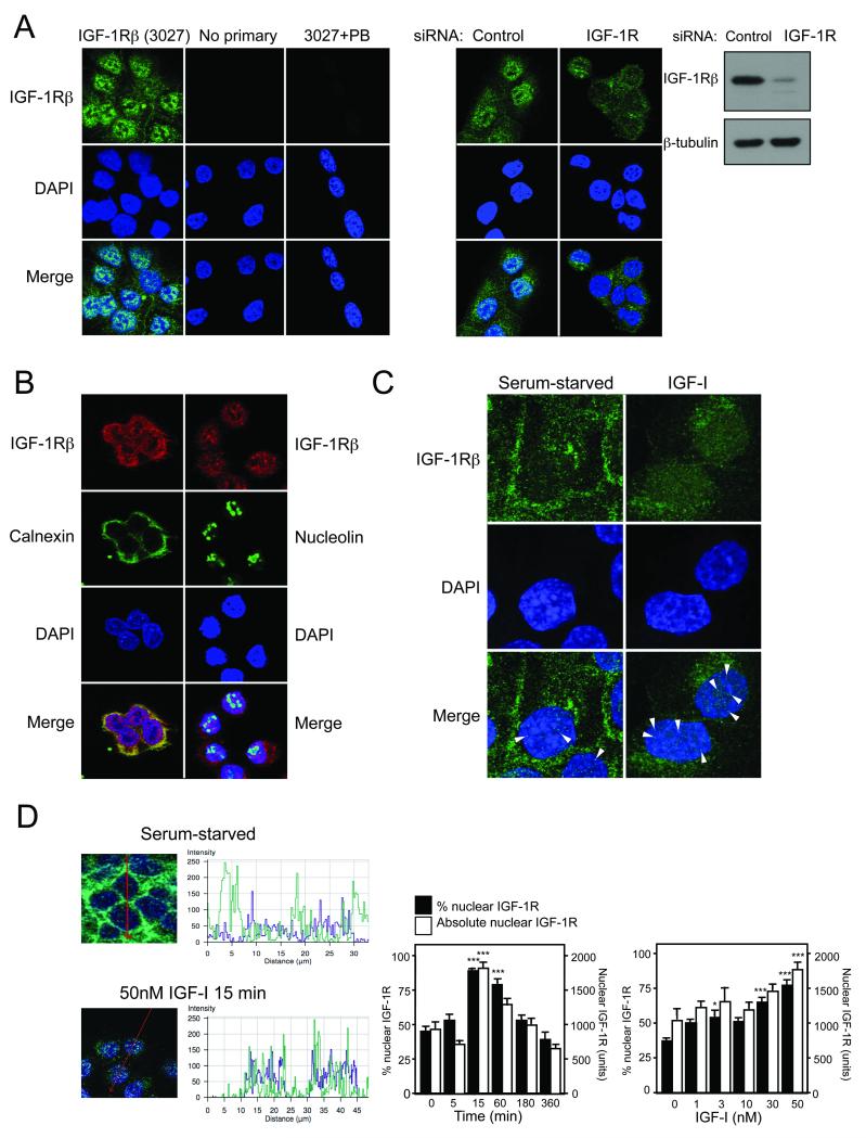
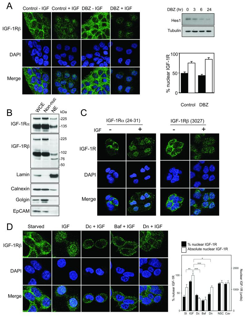
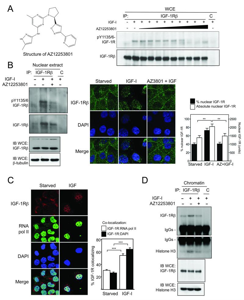
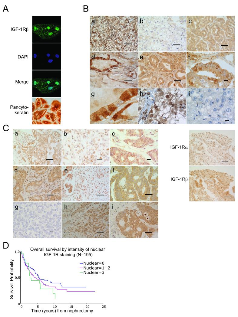
The type 1 insulin-like growth factor receptor (IGF-1R) is a transmembrane glycoprotein composed of two extracellular alpha subunits and two beta subunits with tyrosine kinase activity. The IGF-1R is frequently upregulated in cancers and signals from the cell surface to promote proliferation and cell survival. Recent attention has focused on the IGF-1R as a target for cancer treatment. Here, we report that the nuclei of human tumor cells contain IGF-1R, detectable using multiple antibodies to alpha- and beta-subunit domains. Cell-surface IGF-1R translocates to the nucleus following clathrin-mediated endocytosis, regulated by IGF levels. The IGF-1R is unusual among transmembrane receptors that undergo nuclear import, in that both alpha and beta subunits traffic to the nucleus. Nuclear IGF-1R is phosphorylated in response to ligand and undergoes IGF-induced interaction with chromatin, suggesting direct engagement in transcriptional regulation. The IGF dependence of these phenomena indicates a requirement for the receptor kinase, and indeed, IGF-1R nuclear import and chromatin binding can be blocked by a novel IGF-1R kinase inhibitor. Nuclear IGF-1R is detectable in primary renal cancer cells, formalin-fixed tumors, preinvasive lesions in the breast, and nonmalignant tissues characterized by a high proliferation rate. In clear cell renal cancer, nuclear IGF-1R is associated with adverse prognosis. Our findings suggest that IGF-1R nuclear import has biological significance, may contribute directly to IGF-1R function, and may influence the efficacy of IGF-1R inhibitory drugs.


Figures

Figure 1
Figure 1. IGF-I induces IGF-1R nuclear translocation in human tumor cells
A) IGF-1Rβ immunofluorescence in DU145 cells cultured in complete medium. IGF-1R signal was attenuated by IGF-1R depletion (confirmed in immunoblot to right). B) DU145 cells co-stained for IGF-1R and calnexin or nucleolin. C) Serum-starved DU145 cells were treated with solvent or IGF-I (50nM, 15min), and IGF-1Rβ stained as A. Arrowheads: examples of punctate nuclear IGF1R. Original magnification ×100. D) Left: DU145 cells were serum-starved or IGF-treated (50nM, 15 min), and stained for IGF1Rβ and DAPI. Merged images; arrow shows path along which intensity of IGF-1R (green) and DAPI (blue) is quantified. Center panels: IGF-1R overlying DAPI registers ~50 arbitrary units in starved cells (upper), 100-150 units after IGF-I (lower). Right: quantification of nuclear IGF-1R, following 50nM IGF-I for 0-360min (left), 0-50nM IGF-I for 15min (right). Black columns: mean % nuclear IGF-1R; white: mean absolute nuclear IGF-1R (arbitrary units); bars: SEM (n=20-30 cells). Compared with serum-starved cells, nuclear IGF-1R signal was enhanced by IGF-I (*p<0.05, ***p<0.001).
Figure 2
Figure 2. Full-length IGF-1R α and β subunits undergo nuclear import following clathrin-dependent endocytosis
A) DU145 cells were treated with DBZ (300nM, 6hr) and in final 15min with 50nM IGF-I. Graph: mean % nuclear IGF-1R in serum-starved (black columns) or IGF-treated cells (white); bars: SEM. Immunoblotting (upper right) confirmed DBZ bioactivity in inhibiting expression of Notch target Hes1. B) DU145 whole cell extract (WCE), non-nuclear components (Non-nuc) and nuclear extract (NE) immunoblotted for IGF-1R, lamin (nucleus), calnexin (ER), golgin-84 (Golgi), and EpCAM (plasma membrane). C) Serum-starved DU145 cells were treated with solvent or IGF-I (50nM, 15min) and stained for IGF-1R -α or -β. D) Serum-starved DU145 cells were treated for 4hr with dansylcadaverine (Dc), bafilomycin A1 (Baf), or dynasore (Dn) and in the final 15min with 50nM IGF-I. Absolute nuclear IGF-1R was enhanced by IGF-I (*p<0.01) and inhibited by Dc, Baf and Dn (*p<0.05, ***p<0.001). Figure S5A shows images of IGF-treated cells following caveolin-1 depletion.
Figure 3
Figure 3. IGF-1R nuclear import and chromatin binding are blocked by IGF-1R inhibition
A) Left: structure of AZ12253801. Right: serum-starved DU145 cells treated with 50nM IGF-I in final 15min of 1hr incubation with 0.1-100nM AZ12253801. IGF-1Rβ or control (C) immunoprecipitates probed for phospho- and total IGF-1Rβ Figure S6 shows quantification of these results, and effects on clonogenic survival. B) DU145 cells treated with 50nM IGF-I in final 30min of 6hr incubation with 120nM AZ12253801. Left: nuclear extracts immunoprecipitated with control (C) or IGF-1Rβ antibody, and probed for phospho- and total IGF-1Rβ Right: parallel cultures imaged for IGF-1Rβ. IGF-induced nuclear IGF-1R import was inhibited by AZ12253801 (p<0.001 for % nuclear signal, **p<0.01 for absolute nuclear signal). C) Serum-starved and IGF-treated cells were co-stained for IGF-1Rβ (red) and RNA polymerase II (green). IGF-I enhanced co-localization of IGF-1R with RNA pol II and DAPI (***p<0.001). D) After treatment with AZ12253801 and IGF-I as B), IGF-1Rβ was immunoprecipitated from chromatin and probed for IGF-1Rβ and histone H3.
Figure 4
Figure 4. Nuclear IGF-1R is detectable in human tumors, and is associated with poor prognosis in RCC
A) Detection of nuclear IGF-1Rβ in primary RCC cells. Pancytokeratin positivity confirms epithelial origin. B) Upper: IGF-1Rβ immunohistochemistry in RCC (a-c, h, i) and prostate cancer (d-g) showing heterogeneous staining, with nuclear IGF-1R in a, d, g (high power view of d), h. Scale bar a-e 50μm; f-i 10μm. Lower: Prostate cancer stained for IGF-1R -α (24-31) or -β (3027 CST). Scale bar 50μm. C) Numerous human tumors contain nuclear IGF-1R. a-b: ductal carcinoma of breast, c: DCIS; d: non-small cell lung cancer; e: pancreatic adenocarcinoma; f: colon cancer; g: lymphoma; h: uterine MMMT; i: ovarian serous adenocarcinoma. Nuclear IGF-1Rβ detected in invasive cancers (a, b, d, e, h, i) and DCIS (c). Scale bar a, d-f, h, i 50μm; b, c, g 10μm. D) TMAs containing 195 clear cell RCCs stained for IGF-1Rβ and scored for nuclear IGF-1R intensity: 0 (nil), 1 (light), 2 (moderate), 3 (heavy). Nuclear IGF-1R intensity was associated with adverse prognosis (p=0.005).

References

    1. Chitnis MM, Yuen JS, Protheroe AS, Pollak M, Macaulay VM. The type 1 insulin-like growth factor receptor pathway. Clin Cancer Res. 2008;14:6364–70. - PubMed
    1. Adams TE, Epa VC, Garrett TP, Ward CW. Structure and function of the type 1 insulin-like growth factor receptor. Cell Mol Life Sci. 2000;57:1050–93. - PMC - PubMed
    1. Vecchione A, Marchese A, Henry P, Rotin D, Morrione A. The Grb10/Nedd4 complex regulates ligand-induced ubiquitination and stability of the insulin-like growth factor I receptor. Mol Cell Biol. 2003;23:3363–72. - PMC - PubMed
    1. Romanelli RJ, LeBeau AP, Fulmer CG, Lazzarino DA, Hochberg A, Wood TL. Insulin-like growth factor type-I receptor internalization and recycling mediate the sustained phosphorylation of Akt. J Biol Chem. 2007;282:22513–24. - PubMed
    1. Lin SY, Makino K, Xia W, et al. Nuclear localization of EGF receptor and its potential new role as a transcription factor. Nat Cell Biol. 2001;3:802–8. - PubMed

Publication types

Substances