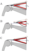
Plasticity of muscle architecture after supraspinatus tears
- PMID: 20710096
- PMCID: PMC4321894
- DOI: 10.2519/jospt.2010.3279
Plasticity of muscle architecture after supraspinatus tears
Abstract
Study design: Controlled laboratory study.
Objectives: To measure the architectural properties of rat supraspinatus muscle after a complete detachment of its distal tendon.
Methods: Supraspinatus muscles were released from the left humerus of 29 Sprague-Dawley rats (mass, 400-450 g), and the animals were returned to cage activity for 2 weeks (n=12), 4 weeks (n=9), or 9 weeks (n=8), before euthanasia. Measurements of muscle mass, pennation angle, fiber bundle length (sarcomere number), and sarcomere length permitted calculation of normalized fiber length, serial sarcomere number, and physiological cross-sectional area.
Results: Coronal oblique sections of the supraspinatus confirmed surgical transection of the supraspinatus muscle at 2 weeks, with reattachment by 4 weeks. Muscle mass and length were significantly lower in released muscles at 2 weeks, 4 weeks, and 9 weeks. Sarcomere lengths in released muscles were significantly shorter at 2 weeks but not different by 4 weeks. Sarcomere number was significantly reduced at 2 and 4 weeks, but returned to control values by 9 weeks. The opposing effects of smaller mass and shorter fibers produced significantly smaller physiological cross-sectional area at 2 weeks, but physiological cross-sectional area returned to control levels by 4 weeks.
Conclusions: Release of the supraspinatus muscle produced early radial and longitudinal atrophy of the muscle. The functional implications of these adaptations would be most profound at early time points (particularly relevant for rehabilitation), when the muscle remains smaller in cross-sectional area and, due to reduced sarcomere number, would be forced to operate over a wider range of the length-tension curve and at higher velocities, all adaptations resulting in compromised force-generating capacity. These data are relevant to physical therapy because they provide tissue-level insights into impaired muscle and shoulder function following rotator cuff injury.
Figures


Similar articles
-
Muscle Architecture Properties of the Deep Region of the Supraspinatus: A Cadaveric Study.Orthop J Sports Med. 2024 Oct 14;12(10):23259671241275522. doi: 10.1177/23259671241275522. eCollection 2024 Oct. Orthop J Sports Med. 2024. PMID: 39421045 Free PMC article.
-
The effect of age on rat rotator cuff muscle architecture.J Shoulder Elbow Surg. 2014 Dec;23(12):1786-1791. doi: 10.1016/j.jse.2014.03.002. Epub 2014 May 28. J Shoulder Elbow Surg. 2014. PMID: 24878035 Free PMC article.
-
Reduced muscle fiber force production and disrupted myofibril architecture in patients with chronic rotator cuff tears.J Shoulder Elbow Surg. 2015 Jan;24(1):111-9. doi: 10.1016/j.jse.2014.06.037. Epub 2014 Sep 3. J Shoulder Elbow Surg. 2015. PMID: 25193488
-
Sarcomere length of torn rotator cuff muscle.J Shoulder Elbow Surg. 2009 Nov-Dec;18(6):955-9. doi: 10.1016/j.jse.2009.03.009. Epub 2009 Jun 10. J Shoulder Elbow Surg. 2009. PMID: 19515583
-
Ibuprofen Differentially Affects Supraspinatus Muscle and Tendon Adaptations to Exercise in a Rat Model.Am J Sports Med. 2016 Sep;44(9):2237-45. doi: 10.1177/0363546516646377. Epub 2016 Jun 8. Am J Sports Med. 2016. PMID: 27281275 Free PMC article.
Cited by
-
Clinical perspectives for repairing rotator cuff injuries with multi-tissue regenerative approaches.J Orthop Translat. 2022 Aug 24;36:91-108. doi: 10.1016/j.jot.2022.06.004. eCollection 2022 Sep. J Orthop Translat. 2022. PMID: 36090820 Free PMC article. Review.
-
Muscle Architecture Properties of the Deep Region of the Supraspinatus: A Cadaveric Study.Orthop J Sports Med. 2024 Oct 14;12(10):23259671241275522. doi: 10.1177/23259671241275522. eCollection 2024 Oct. Orthop J Sports Med. 2024. PMID: 39421045 Free PMC article.
-
Possible involvement of IGF-1 signaling on compensatory growth of the infraspinatus muscle induced by the supraspinatus tendon detachment of rat shoulder.Physiol Rep. 2014 Jan 13;2(1):e00197. doi: 10.1002/phy2.197. eCollection 2014 Jan 1. Physiol Rep. 2014. PMID: 24744876 Free PMC article.
-
Inhibition of p38 mitogen-activated protein kinase signaling reduces fibrosis and lipid accumulation after rotator cuff repair.J Shoulder Elbow Surg. 2016 Sep;25(9):1501-8. doi: 10.1016/j.jse.2016.01.035. Epub 2016 Apr 7. J Shoulder Elbow Surg. 2016. PMID: 27068389 Free PMC article.
-
Tenotomy-induced muscle atrophy is sex-specific and independent of NFκB.Elife. 2022 Dec 12;11:e82016. doi: 10.7554/eLife.82016. Elife. 2022. PMID: 36508247 Free PMC article.
References
-
- Abrams RA, Tsai AM, Watson B, Jamali A, Lieber RL. Skeletal muscle recovery after tenotomy and 7-day delayed muscle length restoration. Muscle & Nerve. 2000;23:707–714. - PubMed
-
- Barton ER, Gimbel JA, Williams GR, Soslowsky LJ. Rat supraspinatus muscle atrophy after tendon detachment. J Orthop Res. 2005;23:259–265. - PubMed
-
- Belozerova IN, Nemirovskaya TL, Shenkman BS, Savina NV, Kozlovskaya IB. Structural and metabolic characteristics of human soleus fibers after long duration spaceflight. J Gravit Physiol. 2002;9:P125–126. - PubMed
-
- Bodine SC, Roy RR, Meadows DA, et al. Architectural, histochemical, and contractile characteristics of a unique biarticular muscle: the cat semitendinosus. J Neurophysiol. 1982;48:192–201. - PubMed
-
- Codman EA. Rupture of the supraspinatus tendon. Clin Orthop. 1990;254:3–26. - PubMed
Publication types
MeSH terms
Grants and funding
LinkOut - more resources
Full Text Sources
Medical

