. 2010 Sep;13(9):1107-12.
doi: 10.1038/nn.2617. Epub 2010 Aug 15.

Rod photoreceptors drive circadian photoentrainment across a wide range of light intensities

Affiliations

Rod photoreceptors drive circadian photoentrainment across a wide range of light intensities

Cara M Altimus et al. Nat Neurosci. 2010 Sep.

Abstract

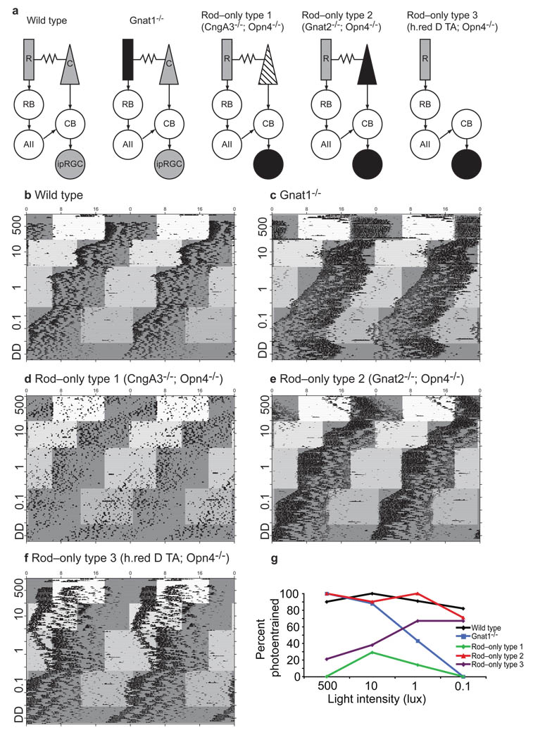
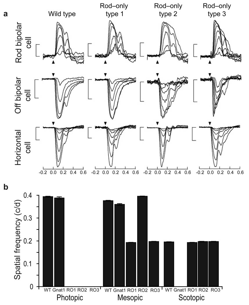
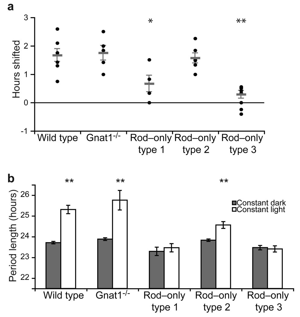
In mammals, synchronization of the circadian pacemaker in the hypothalamus is achieved through direct input from the eyes conveyed by intrinsically photosensitive retinal ganglion cells (ipRGCs). Circadian photoentrainment can be maintained by rod and cone photoreceptors, but their functional contributions and their retinal circuits that impinge on ipRGCs are not well understood. Using mice that lack functional rods or in which rods are the only functional photoreceptors, we found that rods were solely responsible for photoentrainment at scotopic light intensities. Rods were also capable of driving circadian photoentrainment at photopic intensities at which they were incapable of supporting a visually guided behavior. Using mice in which cone photoreceptors were ablated, we found that rods signal through cones at high light intensities, but not at low light intensities. Thus, rods use two distinct retinal circuits to drive ipRGC function to support circadian photoentrainment across a wide range of light intensities.


Figures

Figure 1
Figure 1. Rods drive circadian photoentrainment across a wide range of light intensities
(A) Retinal schematics for all transgenic mouse lines used; gray is functional photoreceptor, black is non–functional resembling dark state, and striped is non–functional resembling saturating light state. (B–F) Representative double–plotted wheel running activity records for (B) WT, (C) Gnat1–/–, (D) Rod–only type 1, (E) Rod–only type 2, and (F) Rod–only type 3 mice assaying for photoentrainment to a 12: 12 hour light: dark cycle which advances six hours concurrently with each intensity decrease. Local time is indicated at the top of each graph and light intensity (lux) is indicated along y–axis of each actogram. Mice were exposed to a 6 hour advanced cycle before the start of this experiment. Note the re–entrainment time course at the 500 lux intensity in all mice which were able to photoentrain (panels B, C, and E, but not D and F). (G) Summary of percentage of photoentrained animals for all genotypes. Refer to Table 1 for number of animals and statistics for each genotype at each light intensity.
Figure 2
Figure 2. Rod–cone pathway is important for mesopic light signaling
(A) Representative traces from current clamp recordings of membrane potential as a function of time from rod bipolar, off bipolar and horizontal cells of WT, rod–only type 1, rod–only type 2, and rod–only type 3 mice show that rod input to each cell type is intact. Arrow represents timing of a 10 ms flash whose strength was increased by a factor of two from generating a just–detectable response to response saturation. Flash strengths for all cells ranged from 0.2 to 30 R* per rod. All scale bars are 10mV. (B) The acuity (cycles/ degree) of mice lacking rod function (Gnat1–/–) is comparable to WT mice at photopic light intensities, while the acuity of rod–only type 1, rod–only type 2, and rod–only type 3 animals is equivalent to that seen in WT animals in scotopic light. Rod–only type 2, but not rod–only types 1 and rod–only type 3, showed similar visual acuity to WT at mesopic light levels. Note that all rod–only mice failed to track at photopic light intensities, while Gnat1–/– animals did not track at scotopic light intensities. Error bars represent SEM.
Figure 3
Figure 3. Rods contribution to phase shifts and period lengthening in constant light is dependent on cone state
(A) WT, Gnat1–/–, and rod–only type 2 mice respond similarly in response to 15 minute, 1000 lux white light pulse administered at CT16 while rod–only type 1 and rod–only type 3 animals show minimal shift in activity onset. (B) Period length in constant darkness is compared to period length in constant light in all animals. WT, Gnat1–/– and rod–only type 2 animals all show significant period lengthening in constant light however, rod–only type 2 period length in constant light in significantly shorter than that seen in WT animals. Rod–only type 1 and 3 animals show no significant period lengthening in constant light. p<0.05 is represented by * and p<0.01 is represented by **. Error bars represent SEM.

References

    1. Guler AD, et al. Melanopsin cells are the principal conduits for rod-cone input to nonimage-forming vision. Nature. 2008;453:102–105. - PMC - PubMed
    1. Hatori M, et al. Inducible ablation of melanopsin-expressing retinal ganglion cells reveals their central role in non-image forming visual responses. PLoS ONE. 2008;3:e2451. - PMC - PubMed
    1. Hattar S, Liao HW, Takao M, Berson DM, Yau KW. Melanopsin-containing retinal ganglion cells: architecture, projections, and intrinsic photosensitivity. Science. 2002;295:1065–1070. - PMC - PubMed
    1. Goz D, et al. Targeted destruction of photosensitive retinal ganglion cells with a saporin conjugate alters the effects of light on mouse circadian rhythms. PLoS ONE. 2008;3:e3153. - PMC - PubMed
    1. Berson DM, Dunn FA, Takao M. Phototransduction by retinal ganglion cells that set the circadian clock. Science. 2002;295:1070–1073. - PubMed

Publication types

MeSH terms