Review
. 2011 Jan;8(1):1-11.
doi: 10.1038/cmi.2010.38. Epub 2010 Aug 16.

Natural killer cell-triggered vascular transformation: maternal care before birth?

Affiliations
Review

Natural killer cell-triggered vascular transformation: maternal care before birth?

Jianhong Zhang et al. Cell Mol Immunol. 2011 Jan.

Abstract

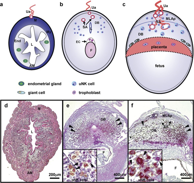
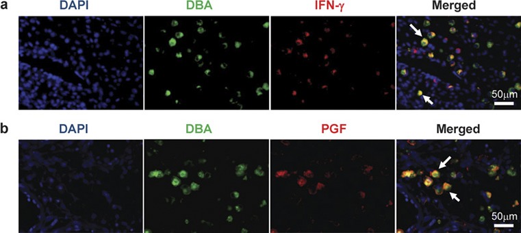
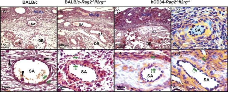
Natural killer (NK) cells are found in lymphoid and non-lymphoid organs. In addition to important roles in immune surveillance, some NK cells contribute to angiogenesis and circulatory regulation. The uterus of early pregnancy is a non-lymphoid organ enriched in NK cells that are specifically recruited to placental attachment sites. In species with invasive hemochorial placentation, these uterine natural killer (uNK) cells, via secretion of cytokines, chemokines, mucins, enzymes and angiogenic growth factors, contribute to the physiological change of mesometrial endometrium into the unique stromal environment called decidua basalis. In humans, uNK cells have the phenotype CD56(bright)CD16(dim) and they appear in great abundance in the late secretory phase of the menstrual cycle and early pregnancy. Gene expression studies indicate that CD56(bright)CD16(dim) uterine and circulating cells are functionally distinct. In humans but not mice or other species with post-implantation decidualization, uNK cells may contribute to blastocyst implantation and are of interest as therapeutic targets in female infertility. Histological and genetic studies in mice first identified triggering of the process of gestation spiral arterial modification as a major uNK cell function, achieved via interferon (IFN)-γ secretion. During spiral arterial modification, branches from the uterine artery that traverse the endometrium/decidua transiently lose their muscular coat and ability to vasoconstrict. The expression of vascular markers changes from arterial to venous as these vessels dilate and become low-resistance, high-volume channels. Full understanding of the vascular interactions of human uNK cells is difficult to obtain because endometrial time-course studies are not possible in pregnant women. Here we briefly review key information concerning uNK cell functions from studies in rodents, summarize highlights concerning human uNK cells and describe our preliminary studies on development of a humanized, pregnant mouse model for in vivo investigations of human uNK cell functions.


Figures

Figure 1
Figure 1
Structure of mouse implantation sites in cross-section. The upper panel drawings illustrate regions of the mouse uterus as virgin (a), gd6–8 (b) and gd10–12 (c). The lower panel presents matched photomicrographs of midsaggital sections of C57BL/6J uterus dually stained with PAS and DBA lectin to detect uNK cells in virgin (d), gd8 (e) and gd10 (f). PAS+DBA+ and PAS+DBA uNK cells, shown in high power insets, appear in mouse DB after embryonic implantation has occurred (arrowheads indicated). Briefly, the uNK cell population's life history in mouse endometrium is summarized as undetectable in virgin uterus (a, d), enriched by the end of first trimester (b, e), proliferating and functional in the second trimester (c, f) and dying in the third trimester (not shown). DBA lectin is useful for uNK cell identification in mice but not in all species. AM, antimesometrial side; DB, decidua basalis; DBA, Dolichos biflorus agglutinin; EC, ectoplacental cone; End, endometrium; F, fetus; gd, gestation day; L, uterine lumen; M, mesometrial side; MLAp, the transient mesometrial lymphoid aggregate of pregnancy; My, myometrium; P, placenta; PAS, periodic acid Schiff; SA, spiral artery; Ua, uterine artery; uNK cell, uterine natural killer cells.
Figure 2
Figure 2
Fluorescence photomicrographs show colocalization of IFN-γ (a) or PGF (b) to uNK cells in gd10 C57BL/6 DB. Mouse uNK cells in 4% PFA fixed, paraffin-embedded tissue were tagged by FITC-conjugated DBA lectin then stained with anti-PGF (Abcam, Cambridge, MA, USA) or anti-IFN-γ (Mebtech, Mariemont, OH, USA), which was visualized by Alexa 594 goat-antirabbit antibody or Alexa Fluor 594-conjugated Streptavidin (Molecular Probes, Eugene, OR, USA). DNA was labeled with DAPI (blue). DBA+PGF+ and DBA+IFN-γ+ uNK cells are indicated (arrows). DAPI, 4,6-diamidino-2-phenylindole; DBA, Dolichos biflorus agglutinin; gd, gestation day; IFN-γ, interferon-γ PFA, paraformaldehyde; PGF, placenta growth factor; uNK cells, uterine natural killer cells.
Figure 3
Figure 3
Fluorescence photomicrographs showing EGFL7 expression at gd6 (a), gd8 (b), gd10 (c) and gd12 (d) in C57BL/6J implantation sites. PFA-fixed paraffin-embedded implantation sites were sectioned, stained with anti-EGFL7 (a kind gift from Dr Huilian Ye, Tumor Biology and Angiogenesis Department, Genentech Inc., South San Francisco, CA, USA), then visualized by PE-conjugated goat anti-Armenian Hamster IgG (red). DNA was labeled with DAPI (blue). EGFL7 had a restricted pattern of expression at the maternal–fetal interface. EGFL7 was transiently expressed at gd10 DB in vein ECs and downregulated by gd12. No uNK cell with detectable EGFL7 was seen. Modified spiral arteries that become vein-like EPHB4+ vessels are shown in gd10–12 implantation sites (e, f). UNK cells express the arterial marker EFNB2 at gd6 and 8 but lose this marker and become EPHB4+ (arrow; f) prior to the switch in expression of these molecules by the spiral arteries. DAPI, 4,6-diamidino-2-phenylindole; EGFL7, epidermal growth factor-like domain 7; gd, gestation day; IgG, immunoglobulin G; PE, phycoerythrin; PFA, paraformaldehyde; uNK cell, uterine natural killer cell.
Figure 4
Figure 4
Implantation sites outcomes after human CD34+ cell inoculation. A total of 16 BALB/c-Rag2/Il2rg/ mice were pretreated with 5-FU (150 mg/kg) and inoculated with 1×10⁵ human CD34+ cord blood cells. Six weeks later, they were paired with syngeneic males and 14 pregnancies were obtained and studied at gd12. (a) The reproductive performance in our colony at gd12 for wild-type BALB/c, BALB/c-Rag2/Il2rg/and human CD34+ cell-inoculated BALB/c-Rag2/Il2rg/mice. There was no significant difference for either viable or total numbers of implantation site between these groups. (b) Human genomic DNA detection by PCR. Genomic DNA was extracted from maternal liver, spleen, and individual MLAps and deciduas of gd12 BALB/c-Rag2/Il2rg/ females using Qiagen QIAamp DNA Blood Mini Kit. The resulting genomic DNA was used as the PCR template. Primer sequences were: human chromosome 17-specific α-satellite (h17α) sequences, 5'-ACACTCTTTTTGCAGGATCTA-3' (forward) and 5'-AGCAATGTGAAACTCTGGGA-3' (reverse); mouse glyceraldehyde-3-phosphate dehydrogenase (mGapdh), 5'-GGTCGGTGTGAACGGATTTGGC-3' (forward) and 5'-GTGGGGTCTCGCTCCTGGAAGA-3' (reverse). PCR was performed under the following conditions: 94°C for 3 min (1 cycle); 94°C for 30 s, 55°C for 30 s, 72°C for 1 min (33 cycles); and 72°C for 10 min (1 cycle). PCR products were separated on 1.0% agarose gels and visualized by ethidium bromide staining. Detection of human chromosome 17-specific α-satellite sequences from an individual pregnancy is shown and verifies that human graft derived cells are present within the implantation site. D, decidual basalis; gd, gestation day; L, liver; M, MLAp (mesometrial lymphoid aggregate of pregnancy); NC, negative control (non-transplanted mouse liver DNA); PC, positive control human placenta; S, spleen; 5-FU, 5-fluorouracil.
Figure 5
Figure 5
Photomicrographs comparing gd12 implantation sites from wild-type BALB/c (A, a), BALB/c-Rag2/Il2rg/ (alymphoid; B, b) and human CD34+ cell-engrafted BALB/c-Rag2/Il2rg/ mice (i.v. injection, fresh cells, 3× IL15/IL15Ra treatment) (C, c1c3). All samples were fixed with 4% PFA and wax embedded using standard histological methods, sectioned at 7 µm then H&E stained. (AC) Low power images of the fetal maternal interface. These lower panels provide high power images of SAs from the same implantation sites. Wild-type SAs are post-modification by gd12 and have widely diluted lumens and very thin walls (a), relative to the lumens and walls of unmodified SA of alymphoid mice (b). Six weeks after inoculation of human CD34+ cord blood cells, pregnant alymphoid mice had abnormal SAs but the extent of abnormality varied greatly between littermates, (C, c1) are from the same implantation site while (c2, c3) are a littermate implantation site. In (C, c1), SA walls were hugely thickened, lacked eosinophilic collagen fibers and contained small pools of red blood cells that appeared to be developing vasculature. In some vessels, individual cells with the same unusual properties were found localized to SA walls (c2, c3). Mononuclear lymphoid cells were not seen (c1c3). In all genotypes, the thickness of the SA wall is indicated by pairs of arrows. UNK cells (arrow head; a) were only identified in wild-type BALB/c where they associated with blood vessels in the DB. DB, decidua basalis; H&E, hematoxylin and eosin; MLAp, mesometrial lymphoid aggregate of pregnancy; DB, decidua basalis, gd, gestation day; iv, intravenous; PFA, paraformaldehyde; SA, spiral artery; uNK cell, uterine natural killer cell.

Similar articles

Cited by

References

    1. Reeves RK, Gillis J, Wong FE, Yu Y, Connole M, Johnson RP. CD16− natural killer cells: enrichment in mucosal and secondary lymphoid tissues and altered function during chronic SIV infection. Blood. 2010;115:4439–4446. - PMC - PubMed
    1. Sun JC, Beilke JN, Lanier LL. Adaptive immune features of natural killer cells. Nature. 2009;457:557–561. - PMC - PubMed
    1. Peel S. Granulated metrial gland cells. Adv Anat Embryol Cell Biol. 1989;115:1–112. - PubMed
    1. Manaster I, Mizrahi S, Goldman-Wohl D, Sela HY, Stern-Ginossar N, Lankry D, et al. Endometrial NK cells are special immature cells that await pregnancy. J Immunol. 2008;181:1869–1876. - PubMed
    1. Lash GE, Robson SC, Bulmer JN. Review: functional role of uterine natural killer (uNK) cells in human early pregnancy decidua. Placenta. 2010;31:S87–S92. - PubMed

Publication types

MeSH terms

Grants and funding