. 2010 Oct 28;29(43):5839-49.
doi: 10.1038/onc.2010.318. Epub 2010 Aug 16.

A Rac-Pak signaling pathway is essential for ErbB2-mediated transformation of human breast epithelial cancer cells

Affiliations

A Rac-Pak signaling pathway is essential for ErbB2-mediated transformation of human breast epithelial cancer cells

L E Arias-Romero et al. Oncogene. .

Abstract

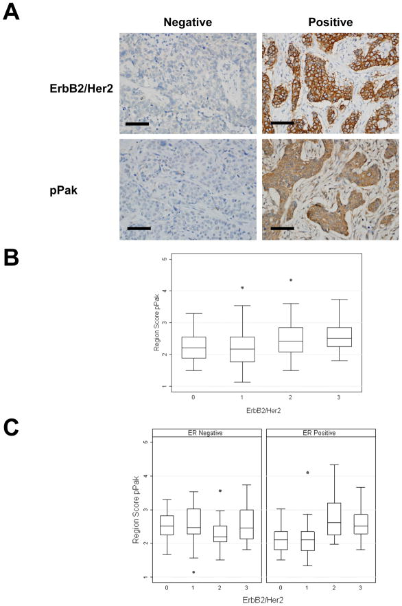
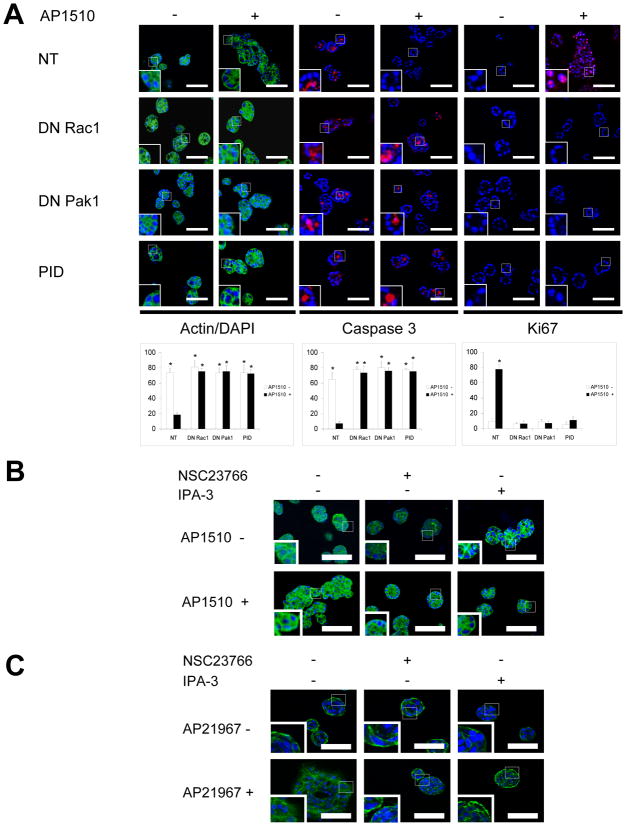
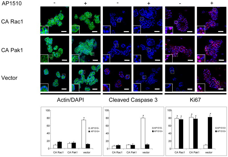
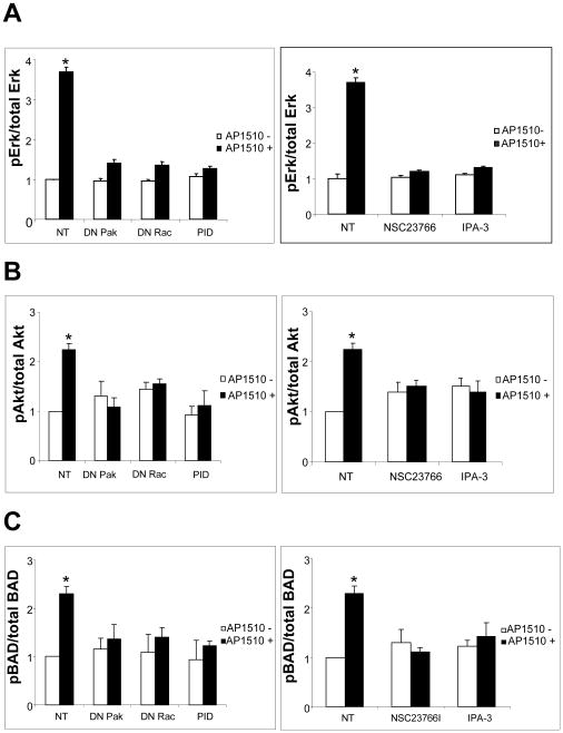
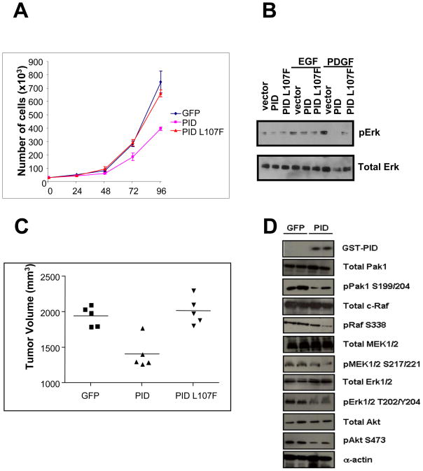
The activation of receptor tyrosine kinases, particularly ErbB2, has an important role in the genesis of breast cancer. ErbB2 kinase activity promotes Ras-mediated stimulation of downstream protein kinase cascades, including the Ras/Raf-1/MAPK/ERK kinase (Mek)/extracellular signal-regulated kinase (Erk) pathway, leading to tumor cell growth and migration. Signaling through the Ras-Erk pathway can be influenced by p21-activated kinase-1 (Pak1), an effector of the Rho family GTPases Rac and Cdc42. In this study, we asked if ErbB2 expression correlates with Pak1 and Erk activity in human breast cancer specimens, and if Pak1 signaling is required for ErbB2 transformation in a three-dimensional (3D) in vitro setting and in xenografts. We found a correlation between ErbB2 expression and activation of Pak in estrogen receptor-positive human breast tumor samples and observed that in 3D cultures, activation of Rac-Pak1 pathway by ErbB2 homodimers induced growth factor-independent proliferation and promoted disruption of 3D mammary acinar-like structures through activation of the Erk and Akt pathways. Further, we found that inhibition of Pak1 by small molecules compromised activation of Erk and Akt, resulting in reversion of the malignant phenotype and restoration of normal acinar architecture. Finally, ErbB2-amplified breast cancer cells expressing a specific Pak inhibitor showed delayed tumor formation and downregulation of Erk and Akt signaling in vivo. These data imply that the Rac-Pak pathway is vital to ErbB2-mediated transformation and that Pak inhibitors represent plausible drug targets in breast cancers in which ErbB2 signaling is activated.


Conflict of interest statement

Conflict of Interest. The authors have no competing financial interests in relation to the work described in this paper.

Figures

Figure 1
Figure 1. Correlation of immunohistochemical staining of ErbB2 and phospho-Pak in human breast cancer
A, Representative example of human breast cancer specimens from TMA that stained positive or negative for ErbB2. Matching specimens from the same patient are shown for phospho-Pak. Size bar = 10 μm. B, TMA-IHC analysis. Correlation of ErbB2 expression with phospho-Pak, the graphic represent the integrated optical density (Region Score) of immunohistochemical staining intensity (r=0.2938, p<0.0001). X axis = ErbB2 staining score (0–3); y axis = P-Pak intensity score (0–4). C, Correlation of ErbB2 expression with phospho-Pak in ER negative and positive samples, the graphics represent the integrated optical density (Region Score) of immunohistochemical staining intensity (r=−0.0428, p=0.7327 and r=0.4342, p<0.0001 respectively)
Figure 2
Figure 2. Pak is required for ErbB2-mediated transformation of MCF-10A cells
A, 10A.ErbB2 cells were transduced with empty adenovirus or adenoviruses encoding DN Rac1 (Rac1 T17N), DN Pak1 (Pak1 K299R), or the PID, plus an adenovirus encoding a tetracycline transactivator, and plated atop reconstituted basement membrane. Cells were stimulated with vehicle or 1 μM AP1510 on day 3 and fixed on day 12 and stained with Oregon green-phalloidin, Ki-67, or anti-cleaved caspase-3. Insets show higher magnification images. Graphics represent the percentage of unilamellar acini, Ki-67-positive, and anti-cleaved caspase-3-positive acini were scored based on assessment of 50 to 60 acini per well, counting cells in four planes of each acinus. *p<0.05 Size bar = 50 μm. B, 10A.ErbB2 cells plated atop reconstituted basement membrane were treated with vehicle, 20 μmol/L NSC23766, or 10 μmol/L IPA-3, plus 1 μmol/L AP1510, on day 3 as indicated, and fixed on day 12. Medium was replaced (with Rac and Pak inhibitors and AP1510) every 3 days. Cells were stained with Oregon green-phalloidin and DAPI. C, 10A.ErbB2/ErbB1 cells plated atop reconstituted basement membrane were treated with vehicle, 20 μmol/L NSC23766, or 10 μmol/L IPA-3, plus 1 μmol/L AP21967, on day 3 as indicated, and fixed on day 12. Cells were stained with Oregon green-phalloidin and DAPI.
Figure 3
Figure 3. Activated Pak bypasses requirement of ErbB2 activity for transformation
10A.ErbB2 cells were transduced with adenoviruses encoding constitutively active (CA) forms of Rac1 (Rac1 G12V) or Pak1 (Pak1 L107F), or a control adenovirus, plus an adenovirus encoding a tetracycline transactivator, and plated atop reconstituted basement membrane. Cells were stimulated with vehicle or AP1510 on day 3 and fixed on day 12 and processed as before. Insets show higher magnification images. Size bar = 50 μm. Graphics represent the percentage of unilamellar acini, Ki-67-positive, and anti-cleaved caspase-3-positive acini were scored based on assessment of 50 to 60 acini per well. *p<0.05.
Figure 4
Figure 4. Pak inhibition down-regulates proliferation signaling pathways in MCF-10A cells
10A.ErbB2 cells were transduced with adenoviruses encoding DN forms of Rac or Pak, or treated with their specific small-molecule inhibitors and plated atop reconstituted basement membrane. Cells were stimulated with vehicle or AP1510 on day 3 and fixed on day 12. The activity of ERK (A), Akt (B), and BAD (C) was assessed by in-cell Western using phospho specific antibodies. *p<0.05
Figure 5
Figure 5. Downregulation of Pak inhibits the tumorigenicity of MDA-MB-631/DYT2 cells in vivo
A, 3 × 10⁴ MDA-MB-631/DYT2 cells stably expressing GFP, PID or PID L107F were plated in triplicate into 12-well plates. Cells were harvested and counted at 0, 24, 48, 72 and 96 h. The data are representative of three independent experiments. Points, mean; bars, SD. B, Immunoblot analysis of ERK activation in MDA-MB-631/DYT2 cells expressing GFP, PID or PID L107F. C, Tumor size distribution in SCID mice. Tumor formation was assessed by injecting 5 × 10⁵ MDA-MB-631/DYT2 cells infected with either empty virus (GFP), PID or PID L107F into the flanks of the mice. Five mice were used in total for each group. The size of tumors was measured three weeks after injection. Horizontal lines, average tumor diameters. D, Western analysis shows expression and activation levels of the indicated proteins in primary tumor lysates and quantification of relative difference in activation between GFP and PID expressing tumors.

Similar articles

Cited by

References

    1. Amundadottir LT, Leder P. Oncogene. 1998;16:737–746. - PubMed
    1. Deacon SW, Beeser A, Fukui JA, Rennefahrt UE, Myers C, Chernoff J, Peterson JR. Chem Biol. 2008;15:322–331. - PMC - PubMed
    1. Dummler B, Ohshiro K, Kumar R, Field J. Cancer Metastasis Rev. 2009;28:51–63. - PMC - PubMed
    1. Gao Y, Dickerson JB, Guo F, Zheng J, Zheng Y. Proc Natl Acad Sci U S A. 2004;101:7618–7623. - PMC - PubMed
    1. Hardy S, Kitamura M, Harris-Stansil T, Dai Y, Phipps ML. J Virol. 1997;71:1842–1849. - PMC - PubMed

Publication types

MeSH terms