

A case of clear cell eccrine porocarcinoma
- PMID: 20711273
- PMCID: PMC2917690
- DOI: 10.5021/ad.2010.22.3.330
A case of clear cell eccrine porocarcinoma
Abstract
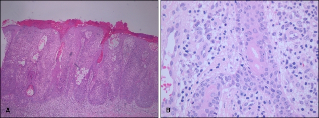
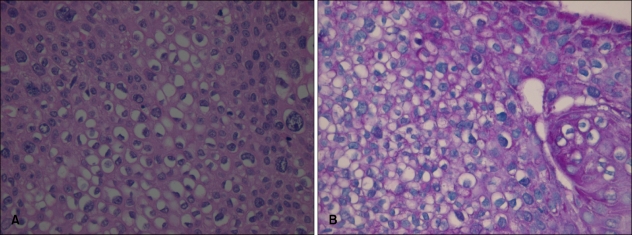
Eccrine porocarcinoma (EP) is a rare malignant tumor arising from the intraepidermal eccrine duct. The tumor cells frequently contain glycogen, but prominent clear cell changes in EP are rarely reported. A 78-year-old woman presented with a slightly pruritic, erythematous, verrucous plaque on her left thigh. Histopathological examination revealed intraepidermal tumor cell nests composed of small basaloid cells and duct-like structures lined by periodic acid-Schiff (PAS)-positive cuticles. Besides the typical findings of EP, clear cell changes were predominantly observed in the tumor cell aggregations. Herein we report a case of the clear cell variant of EP rarely reported in previous literature.
Keywords: Clear cell; Eccrine porocarcinoma; Malignant eccrine poroma.
Figures


References
-
- Rutten A, Requena L, Requena C. Clear-cell porocarcinoma in situ: a cytologic variant of porocarcinoma in situ. Am J Dermatopathol. 2002;24:67–71. - PubMed
-
- Pinkus H, Mehregan AH. Epidermotropic eccrine carcinoma. A case combining features of eccrine poroma and Paget's dermatosis. Arch Dermatol. 1963;88:597–606. - PubMed
-
- Robson A, Greene J, Ansari N, Kim B, Seed PT, McKee PH, et al. Eccrine porocarcinoma (malignant eccrine poroma): a clinicopathologic study of 69 cases. Am J Surg Pathol. 2001;25:710–720. - PubMed
-
- Requena L, Sarasa JL, Pique E, Farina MC, Olivares M, Martin L. Clear-cell porocarcinoma: another cutaneous marker of diabetes mellitus. Am J Dermatopathol. 1997;19:540–544. - PubMed
-
- Saitoh A, Ohtake N, Fukuda S, Tamaki K. Clear cells of eccrine glands in a patient with clear cell syringoma associated with diabetes mellitus. Am J Dermatopathol. 1993;15:166–168. - PubMed
Publication types
LinkOut - more resources
Full Text Sources
