. 2010 Aug 12;5(8):e12109.
doi: 10.1371/journal.pone.0012109.

Positive feedback regulation between phospholipase D and Wnt signaling promotes Wnt-driven anchorage-independent growth of colorectal cancer cells

Affiliations

Positive feedback regulation between phospholipase D and Wnt signaling promotes Wnt-driven anchorage-independent growth of colorectal cancer cells

Dong Woo Kang et al. PLoS One. .

Abstract

Background: Aberrant activation of the canonical Wnt/beta-catenin pathway occurs in almost all colorectal cancers and contributes to their growth, invasion and survival. Phopholipase D (PLD) has been implicated in progression of colorectal carcinoma However, an understanding of the targets and regulation of this important pathway remains incomplete and besides, relationship between Wnt signaling and PLD is not known.

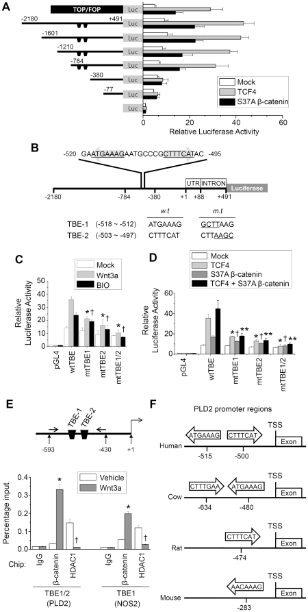
Methodology/principal findings: Here, we demonstrate that PLD isozymes, PLD1 and PLD2 are direct targets and positive feedback regulators of the Wnt/beta-catenin signaling. Wnt3a and Wnt mimetics significantly enhanced the expression of PLDs at a transcriptional level in HCT116 colorectal cancer cells, whereas silencing of beta-catenin gene expression or utilization of a dominant negative form of T cell factor-4 (TCF-4) inhibited expression of PLDs. Moreover, both PLD1 and PLD2 were highly induced in colon, liver and stomach tissues of mice after injection of LiCl, a Wnt mimetic. Wnt3a stimulated formation of the beta-catenin/TCF complexes to two functional TCF-4-binding elements within the PLD2 promoter as assessed by chromatin immunoprecipitation assay. Suppressing PLD using gene silencing or selective inhibitor blocked the ability of beta-catenin to transcriptionally activate PLD and other Wnt target genes by preventing formation of the beta-catenin/TCF-4 complex, whereas tactics to elevate intracellular levels of phosphatidic acid, the product of PLD activity, enhanced these effects. Here we show that PLD is necessary for Wnt3a-driven invasion and anchorage-independent growth of colon cancer cells.

Conclusion/significance: PLD isozyme acts as a novel transcriptional target and positive feedback regulator of Wnt signaling, and then promotes Wnt-driven anchorage-independent growth of colorectal cancer cells. We propose that therapeutic interventions targeting PLD may confer a clinical benefit in Wnt/beta-catenin-driven malignancies.


Conflict of interest statement

Competing Interests: The authors have declared that no competing interests exist.

Figures

Figure 1
Figure 1. Wnt3a and GSK3β inhibitors increase expression of PLD isozymes in HCT116 colon cancer cells.
(A) HCT116 cells were treated with purified recombinant Wnt3a (150 ng/ml) for the indicated times, and the lysates were immunoprecipitated and immunoblotted with antibody to PLD. β-catenin was analyzed by Western blotting using nuclear lysates (upper panel). Histograms show relative protein levels of PLD1 and PLD2, which are normalized to the corresponding α-tubulin values (lower panel). (B-C) HCT116 cells were treated with Wnt3a (150 ng/ml), LiCl (20 mM), or BIO (1 µM) for 24 h. (B) Protein level of PLDs was analyzed by immunoprecipitation and immunoblotting. c-Myc and NOS2 expression was analyzed by Western blotting (upper panel). Histograms show relative protein levels of PLD1 and PLD2, which are normalized to the corresponding α-tubulin values (lower panel). Data are representative of three independent experiments. (C) mRNA levels of the indicated genes were analyzed by Q-RT-PCR. *P<0.05 versus vehicle. (D) The indicated promoter reporter plasmids were transfected and treated with Wnt3a, LiCl, or BIO for 12 h; luciferase activity was then determined. *P<0.01 versus vehicle. Data represent the mean ± S.D. of three independent experiments.
Figure 2
Figure 2. LiCl increases expression of PLD isozymes in vivo.
(A) Mice were intravenously injected with LiCl, as described in “Materials and Methods”. Lysates from various tissues were immunoprecipitated and immunoblotted with antibody to PLD recognizing both PLD1 and PLD2 (left panel). Protein levels were analyzed by immunoblot using the indicated antibodies. Histograms show relative protein levels of PLD1 and PLD2, which are normalized to the corresponding α-tubulin values (right panel). (B) Paraffin sections of colon tissues were subjected to immunofluorescence analyses using anti-β-catenin (Alexa fluor 488; green) and PLD (Alexa fluor 555; red) antibody. Tissues were monitored using Zeiss LSM 510 confocal microscope. Microscopy fields were observed at × 650 magnification. Data are representative of three independent experiments.
Figure 3
Figure 3. β-catenin and TCF-4 enhance the promoter activities and protein levels of PLD isozymes.
(A) HCT116 cells were co-transfected with pGL4-PLD or TOP/FOP reporters and the indicated expression vectors. *P<0.01 versus mock; †P<0.05 versus S37A β-catenin/TCF-4. (B) Cells were transfected with either the TCF-4 or β-catenin expression vector, and lysates were immunoprecipitated or immunoblotted with the indicated antibodies (upper panel). Histograms show relative protein levels of PLD1 and PLD2, which are normalized to the corresponding α-tubulin values (lower panel). (C) Cells were transfected with ΔN30 TCF-4 or shRNA for β-catenin. Lysates were analyzed by immunoprecipitation or Western blot using the indicated antibodies (upper panel). Protein expression was quantitated by densitometer analysis. Histograms show relative protein levels of PLD1 and PLD2, which are normalized to the corresponding α-tubulin values (lower panel). Data are representative of three independent experiments.
Figure 4
Figure 4. β-catenin/TCF-4 specifically binds to the TBEs of the PLD2 promoter and enhances PLD2 expression.
(A) Deletion analysis of pGL4-PLD2 in HCT116 cells. A schematic representation of pGL4-PLD2 reporter constructs is shown. Cells were cotransfected with pGL4-PLD2 and the indicated expression vectors, followed by determination of luciferase activity. (B) Diagrammatic representation of the −2180 to +491 region of the human PLD2 promoter. Numbers above the lines refer to the transcription start site of the PLD2 gene (+1). Two putative binding sites for TCF-4 are indicated on the sequence (the arrows indicate the direction). (C) HCT 116 cells were transfected with the luciferase reporter plasmid containing the wild type (wt) PLD2 promoter, one or double TBE mutant forms (mt) of PLD2 promoter, and treated with Wnt3a (150 ng/ml) or BIO (1 µM). Luciferase activities were measured. *P<0.05 versus wtTBE/Wnt3a; †P<0.01 versus wtTBE/BIO. (D) Cells were co-transfected with the indicated expression vectors, along with the wt or TBE mutant forms of the PLD2 promoter. Luciferase activities were measured. *P<0.05 versus transfected with wtTBE/β-catenin; †P<0.05 versus wtTBE/TCF4; **P<0.05 versus transfected with wtTBE/β-catenin/TCF4. (E) Arrows indicate position of primers used in the ChIP experiment. The ChIP assay was performed using preimmune IgG, anti-β-catenin, or anti-HDAC1 antibody and analyzed by Q-RT-PCR. As a positive control, ChIP analysis of the NOS2 promoter containing TBE was performed. *P<0.05 versus β-catenin/vehicle; †P<0.05 versus HDAC1/vehicle. Data are representative of four independent experiments. (F) Schematic diagram for comparison of TBEs on PLD2 promoter regions from various species. TCF-4 binding elements in 5′ flanking regions of the human PLD2 transcriptional start site (TSS) were compared with those of the genomes from 4 different species. A core motif, CTTTG(A/T)(A/T) [or the complementary sequence (A/T)(A/T)CAAAG] of TCF binding sequences on PLD2 promoter is highly conserved across species.
Figure 5
Figure 5. PLD isozyme is required for formation of the β-catenin/TCF-4 complex and promotion of β-catenin/TCF transcriptional activity.
(A) After HCT116 cells were co-transfected with TOP/FOP reporters, S37A β-catenin, or siRNA for PLDs, cells were treated with or without the indicated dose of VU0155069 or VU0285655-1; TCF activity was then determined. *P<0.05 versus Mock; **P<0.05 versus S37A β-catenin; †P<0.05 versus S37A β-catenin-siRNA. HCT116 cells were transfected with siRNAs for PLD (B) or pretreated with 10 μM of VU0155069 or VU0285655-1 (C), and then stimulated with Wnt3a (150 ng/ml) for 20 h. (D) HCT116 cells were labeled with [3H] myristate and treated with Wnt3a (150 ng/ml) for 12 h. PLD activity was measured as dsescribed in Materials and Methods. *P<0.05 versus vehicle. (E) HCT116 cells were treated with the indicated dose of PA or 1-propranolol for 24 h. Association of TCF-4 with β-catenin was analyzed by immunoprecipitation and immunoblot using the indicated antibodies. Protein levels were determined by immunoprecipitation or immunoblotting using the indicated antibodies. Interaction levels or protein expression were quantitated by densitometer analysis. Data are representative of three independent experiments.
Figure 6
Figure 6. PLD isozyme mediates anchorage-independent growth, migration, and invasion in concert with the Wnt/β-catenin/TCF-dependent pathway.
(A–B) HCT116 cells were transfected with or without siRNA for PLD1 or PLD2, and then seeded in Matrigel-coated invasion chambers or migration chambers and stimulated with purified recombinant Wnt3a (150 ng/ml) for 24 h. The extent of invasion (A) and migration (B) were expressed as an average number of cells per microscopic field. *P<0.01 versus vehicle; †P<0.05 versus Wnt3a/control-siRNA. (C) HCT116 cells were transfected with siRNA for PLD1 or PLD2, suspended in agar matrix, and treated with or without 10 µM of VU0155069 or VU0285655-1 and treated with Wnt3a (150 ng/ml). Following 10 days incubation, the anchorage-independent growth assay was performed as described in the Materials and Methods section. *P<0.05 versus Mock; **P<0.05 versus Wnt3a; †P<0.05 versus Wnt3a/control-siRNA. (D) PLD1 or PLD2 mediates anchorage-independent growth via Wnt signaling. HCT116 cells were transfected with the indicated expression vectors and an anchorage-independent growth assay was performed. *P<0.05 versus Mock; †P<0.05 versus PLD1; ††P<0.05 versus PLD2. Data are representative of three independent experiments.

Similar articles

Cited by

References

    1. Korinek V, Barker N, Morin PJ, van Wichen D, de Weger R, et al. Constitutive transcriptional activation by a beta-catenin-Tcf complex in APC-/- colon carcinoma. Science. 1997;275:1784–1787. - PubMed
    1. Morin PJ. β-Catenin signaling and cancer. Bioassays. 1999;21:1021–1030. - PubMed
    1. Morin PJ, Sparks AB, Korinek V, Barker N, Clevers H, et al. Activation of beta-catenin-Tcf signaling in colon cancer by mutations in beta-catenin or APC. Science. 1997;275:1787–1790. - PubMed
    1. Aberle H, Bauer A, Stappert J, Kispert A, Kemler R. β-catenin is a target for the ubiquitin-proteasome pathway. EMBO J. 1997;16:3797–3804. - PMC - PubMed
    1. Hsu SC, Galceran J, Grosschedl R. Modulation of transcriptional regulation by LEF-1 in response to Wnt-1 signaling and association with beta-catenin. Mol Cell Biol. 1998;18:4807–4818. - PMC - PubMed

Publication types

MeSH terms