. 2010 Aug 12;5(8):e12119.
doi: 10.1371/journal.pone.0012119.

Quantitative organization of GABAergic synapses in the molecular layer of the mouse cerebellar cortex

Affiliations

Quantitative organization of GABAergic synapses in the molecular layer of the mouse cerebellar cortex

Federica Briatore et al. PLoS One. .

Abstract

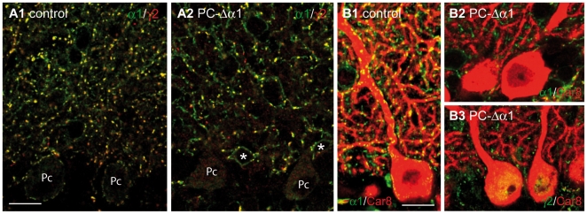
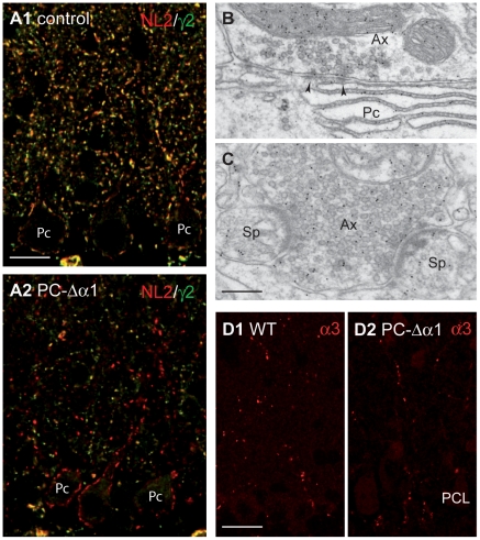
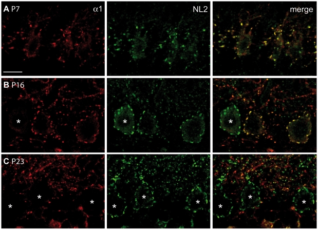
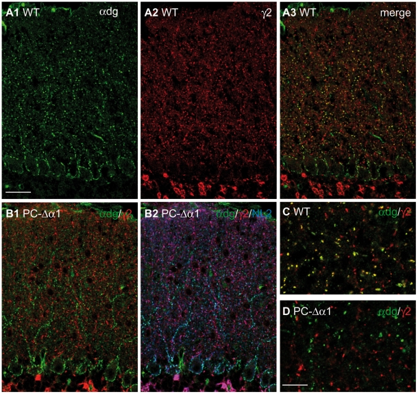
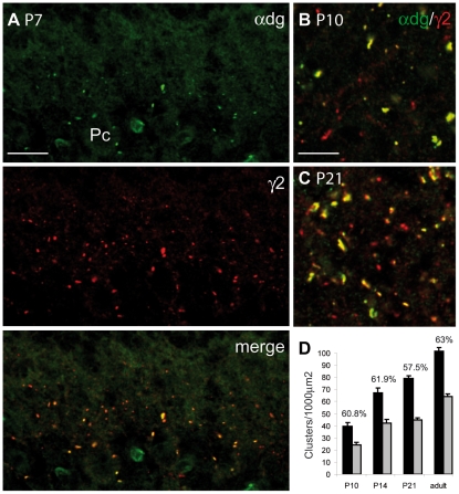
In the cerebellar cortex, interneurons of the molecular layer (stellate and basket cells) provide GABAergic input to Purkinje cells, as well as to each other and possibly to other interneurons. GABAergic inhibition in the molecular layer has mainly been investigated at the interneuron to Purkinje cell synapse. In this study, we used complementary subtractive strategies to quantitatively assess the ratio of GABAergic synapses on Purkinje cell dendrites versus those on interneurons. We generated a mouse model in which the GABAA receptor alpha1 subunit (GABAARalpha1) was selectively removed from Purkinje cells using the Cre/loxP system. Deletion of the alpha1 subunit resulted in a complete loss of GABAAR aggregates from Purkinje cells, allowing us to determine the density of GABAAR clusters in interneurons. In a complementary approach, we determined the density of GABA synapses impinging on Purkinje cells using alpha-dystroglycan as a specific marker of inhibitory postsynaptic sites. Combining these inverse approaches, we found that synapses received by interneurons represent approximately 40% of all GABAergic synapses in the molecular layer. Notably, this proportion was stable during postnatal development, indicating synchronized synaptogenesis. Based on the pure quantity of GABAergic synapses onto interneurons, we propose that mutual inhibition must play an important, yet largely neglected, computational role in the cerebellar cortex.


Conflict of interest statement

Competing Interests: The authors have declared that no competing interests exist.

Figures

Figure 1
Figure 1. Loss of GABAARs from Purkinje cells of PC-Δα1 mice.
(A1,A2) Confocal images showing the distribution of GABAARα1 (green) and GABAARγ2 (red) in the cerebellar cortex of control (A1) and PC-Δα1 mice (A2). Note that in both conditions most GABAARγ2-positive clusters colocalize with the α1 subunit. A few clusters labelled for GABAARγ2 but not for GABAARα1 likely represent synapses containing α3-GABAARs. No surface labelling is visible in Purkinje cells (Pc) of PC-Δα1 mice, whereas MLIs (asterisks) are recognized by extrasynaptic labelling of the α1 subunit (A2). Double-labelled clusters (yellow) identify postsynaptic GABAAR aggregates on the cell body and the dendrites of MLIs (A2). (B1) Clustered distribution of GABAARα1 (green) in control Purkinje cells labelled for Car8 (red). (B2,B3) GABAAR clusters (B2: GABAARα1; B3: GABAARγ2) are not visible in Purkinje cells of PC-Δα1 mice. Scale bars: 15 µm.
Figure 2
Figure 2. Synaptic organization in the cerebellum of PC-Δα1 mice.
(A1,A2) Double labelling for GABAARγ2 (green) and NL2 (red) in control (A1) and PC-Δα1 mice (A2). NL2 colocalizes extensively with the γ2 subunit and also clusters at postsynaptic sites lacking GABAARs in Purkinje cells of PC-Δα1 mice. (B,C) Electron micrographs of the ML of a PC-Δα1 mouse showing that GABA-immunopositive axon terminals (Ax) make both conventional, symmetric synapses (B, arrowheads) with Purkinje cell dendrites (Pc) and heterologous synapses (C) with spines (Sp). (D1,D2) Similar distribution of α3-GABAARs in the ML of control (D1) and PC-Δα1 mice (D2). PCL, Purkinje cell layer. Scale bars: A = 15 µm. B,C = 200 nm. D = 20 µm.
Figure 3
Figure 3. Ablation of GABAARs is protracted during postnatal development and is asynchronous among different Purkinje cells.
Double labelling for GABAARα1 (red) and NL2 (green) in the cerebellar cortex of P7 (A), P16 (B) and P23 (C) PC-Δα1 mice. All P7 Purkinje cells express postsynaptic GABAAR clusters, colocalized with NL2 (A). Purkinje cells labelled for NL2 but not GABAARs (asterisks) are visible at P16 (B). By P23, practically all Purkinje cells are immunonegative for GABAARs (C). Scale bar: 20 µm.
Figure 4
Figure 4. Dystroglycan is present at GABAergic synapses in Purkinje cells but not in cerebellar interneurons.
(A1–A3) Double labelling for α-dystroglycan (green) and GABAARγ2 (red) in the cerebellar cortex of a WT mouse. Labelling for α-dystroglycan outlines the cell bodies and major dendrites of Purkinje cells and colocalizes precisely with GABAARγ2. Labelling for the γ2 subunit is weaker at perisomatic synapses, scarcely visible in these low magnification images. (B1,B2) Triple labelling for α-dystroglycan (green), GABAARγ2 (red) and NL2 (blue) in the cerebellar cortex of a PC-Δα1 mouse. Dystroglycan colocalizes with NL2 exclusively at silent synapses that lack GABAARs (B2, cyan). NL2 associates with GABAARs at interneuron-interneuron synapses, where immunolabelling for α-dystroglycan is not visible (B2, magenta). (C,D) High-magnification images of the ML showing that in WT mice a subset of GABAergic synapses contain α-dystroglycan (yellow clusters), whereas in PC-Δα1 mice α-dystroglycan never colocalizes with GABAARγ2-positive clusters. Scale bars: A,B = 30 µm. C,D = 10 µm.
Figure 5
Figure 5. Early synaptic localization of α-dystroglycan and postnatal development of GABAergic synapses in the ML.
(A) Double labelling for α-dystroglycan and GABAARγ2 in the cerebellar cortex of a P7 wild-type mouse showing that α-dystroglycan clusters at developing GABAergic synapses onto Purkinje cells (Pc). (B,C) Representative images showing that α-dystroglycan associates with a subset of GABAARγ2-positive synapses in the ML of P10 (B) and P21 (C) mice. (D) Synapses were quantified in the iML by counting clusters immunopositive for GABAARγ2 (black bars) and α-dystroglycan (grey bars). Percentage values express the ratio of α-dystroglycan-positive clusters over the entire population of GABA synapses labelled for GABAARγ2 (n = 3 mice for each developmental stage). Note that the values vary little during postnatal development. Scale bars: A = 15 µm. B,C = 6 µm.

References

    1. Ramón y Cajal S. Paris: Maloine; 1911. Histologie du système nerveux de l'homme et des vertébrés, vol.II.
    1. Fox FA, Hillman DE, Sugesmund KA, Dutta CR. The primate cerebellar cortex: A Golgi and electron microscope study. Prog Brain Res. 1967;25:174–225. - PubMed
    1. Palay S, Chan-Palay V. Berlin: Springer; 1974. Cerebellar Cortex: Cytology and Organization.
    1. Eccles J, Ito M, Szentagothai J. Berlin: Springer; 1967. The Cerebellum as a Neuronal Machine.
    1. Marr D. A theory of cerebellar cortex. J Physiol. 1969;202:437–470. - PMC - PubMed

Publication types

LinkOut - more resources