. 2010 Aug 10;8(8):e1000450.
doi: 10.1371/journal.pbio.1000450.

Widespread protein aggregation as an inherent part of aging in C. elegans

Affiliations

Widespread protein aggregation as an inherent part of aging in C. elegans

Della C David et al. PLoS Biol. .

Abstract

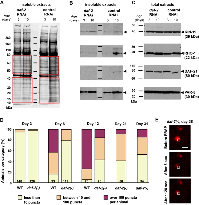
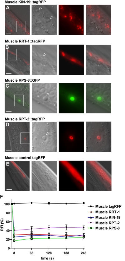
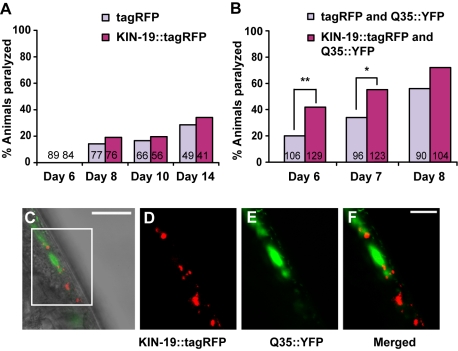
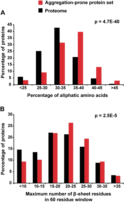
Aberrant protein aggregation is a hallmark of many age-related diseases, yet little is known about whether proteins aggregate with age in a non-disease setting. Using a systematic proteomics approach, we identified several hundred proteins that become more insoluble with age in the multicellular organism Caenorhabditis elegans. These proteins are predicted to be significantly enriched in beta-sheets, which promote disease protein aggregation. Strikingly, these insoluble proteins are highly over-represented in aggregates found in human neurodegeneration. We examined several of these proteins in vivo and confirmed their propensity to aggregate with age. Different proteins aggregated in different tissues and cellular compartments. Protein insolubility and aggregation were significantly delayed or even halted by reduced insulin/IGF-1-signaling, which also slows aging. We found a significant overlap between proteins that become insoluble and proteins that influence lifespan and/or polyglutamine-repeat aggregation. Moreover, overexpressing one aggregating protein enhanced polyglutamine-repeat pathology. Together our findings indicate that widespread protein insolubility and aggregation is an inherent part of aging and that it may influence both lifespan and neurodegenerative disease.


Conflict of interest statement

The authors have declared that no competing interests exist.

Figures

Figure 1
Figure 1. The majority of insoluble-prone proteins are consistently more insoluble with age in C. elegans.
(A) Total and detergent-insoluble protein staining of gon-2(−)/gonad-less extracts using Sypro Ruby. Total protein fraction was diluted 1∶3 compared to the insoluble fraction. Quantification of insoluble proteins in the red outlined areas revealed a fold change of 3.5±0.8 (SD) on average between young and aged animals (two biological and two experimental repeats). (B) Distribution of the fold change shown in logarithm base 2 in levels of insoluble proteins identified in aged animals (when half the population is alive) compared to young animals (Day 3 of adulthood at 25°C). Each dot represents the fold change for one insoluble-prone protein. Red bar indicates the mean. The fold changes of proteins that were identified in both independent experiments are shown for each strain in Experiment 1 and Experiment 2. These results show that proteins identified in the formic acid soluble fraction have a tendency to accumulate with age. 711 proteins were present in all four samples (defined as having an iTRAQ peak above 25 counts in young and/or in old animals) (Experiment 1 and Experiment 2). Of these, some were excluded from the fold change calculations because their iTRAQ peak was too low (≤25 counts) in young animals. Fold changes were calculated in Experiment 1 for 698 of 711 insoluble proteins in fem-1(−) animals and 670 of 711 insoluble proteins in gon-2(−) animals and in Experiment 2 for 692 of 711 insoluble proteins in fem-1(−) and 695 of 711 insoluble proteins in glp-1(−). (C–D) Changes in insolubility with age in the two independent biological replicates were strongly correlated. Age-dependent insolubility fold changes are plotted for both Experiments 1 and 2, comparing fem-1(−) animals (C) and comparing gon-2(−) to glp-1(−) animals (D). Spearman r correlation and two-tailed p values were calculated for each set: fem-(−): r = 0.81, p<0.0001; gon-2(−)/glp-1(−): r = 0.63, p<0.0001.
Figure 2
Figure 2. Age-dependent protein aggregation can occur in different tissues.
(A–B) Ppie-1::gfp::rho-1-expressing animal, Day 1 (A) and Day 9 (B–C). In our proteomic analysis, we identified RHO-1 as a protein prone to aggregate with age both in the reproductive and somatic tissues. GFP::RHO-1, expressed in the germline, was localized to germline stem cell (open arrowhead) and oocyte (full arrowhead) membranes in young animals. With age, GFP::RHO-1 also accumulated in sclerotic oocytes in the uterus (full arrowhead). Scale bar: 20 µm. (D) FRAP-immobile GFP::RHO-1 in aged oocytes. GFP::RHO-1 is pseudocolored in magenta. Laser setting: 25% in 0.85 µm2 (white open box). Scale bar: 2 µm. (E) Quantification of relative fluorescence intensity (RFI) during recovery. We found no recovery of GFP::RHO-1 in sclerotic oocytes in aged animals (Day 12, N animals = 5, N puncta evaluated = 5) but rapid fluorescence recovery for GFP::RHO-1 localized to the germ-line cell membrane in young animals (t1/2 = 35 s, Day 1, N animals = 5, N puncta evaluated = 5) and in aged animals (t1/2 = 17 s, Day 12, N animals = 3, N puncta evaluated = 4). (F–K) Formation of KIN-19::tagRFP puncta in the anterior pharyngeal bulb (metacorpus) with age. Pkin-19::kin-19::tagrfp animals (F and G); daf-2(e1370); Pkin-19::kin-19::tagrfp animals (H); control Pkin-19::tagrfp animals (I and J). (F–J) 10 ms exposure, 100×. (K) Schematic of C. elegans' pharynx, boxed area is shown in F–J. (L) FRAP-immobile KIN-19::tagRFP puncta in the anterior pharyngeal bulb of 15-day-old Pkin-19::kin-19::tagrfp animal. Laser settings: 40% in 0.46 µm2 (white open box). Scale bar: 1 µm. (M) We found no fluorescence recovery of pharyngeal or BDU neuronal KIN-19::tagRFP in aged animals (Day 12, N animals = 5, N puncta evaluated = 5 and Day 12 and 14, N animals = 5, N puncta evaluated = 5, respectively). KIN-19::tagRFP was able to diffuse back at a slow rate in young animals both in the pharynx and neurons (t1/2 = 107 s, Day 1, N animals = 4, N puncta evaluated = 5 and t1/2 = 151 s, Day 3, N animals = 5, N puncta evaluated = 5, respectively). The few puncta formed by tagRFP alone contained highly mobile protein in aged animals (Day 12; N animals = 5, N puncta evaluated = 5).
Figure 3
Figure 3. Insoluble but not total levels of four aggregation-prone proteins increased with age.
(A and C) Western blot detection of KIN-19 (CK1α), RHO-1, DAF-21 (Hsp90), and PAR-5 (14-3-3) in young and aged animals containing either somatic and germline tissue [fem-1(−), (A)] or containing only somatic tissues [gon-2(−), (C)]. The total fraction (Urea and SDS buffer) contains all proteins and the detergent-insoluble fraction contains aggregation-prone proteins. The total protein fraction was diluted 1∶3 compared to insoluble fraction. Arrowheads mark the protein bands corresponding to the aggregation-prone candidates. Overall, Western blot analysis confirms our mass spectrometry results demonstrating a large increase in insolubility with age. With age, we noted a slight decrease in the size of full-length DAF-21 (less than 10 kDa). (B and D) Quantification of the fractional increase in aggregated levels compared to total levels of each candidate evaluated by Western blot. These results demonstrate that age-dependent insolubility for each of the four proteins we examined occurs independently of an increase in total protein levels. Extracts from two biologically independent experiments were evaluated. Error bars indicate SEM.
Figure 4
Figure 4. Reducing KIN-19::tagRFP levels does not prevent age-dependent protein aggregation in the pharynx.
(A) The activity of the kin-19 promoter was up-regulated with age. Fluorescence from the tagRFP reporter driven by the kin-19 promoter increased by 1.6-fold between Day 2 and Day 6 in Pkin-19::tagrfp animals (Unpaired t test *p = 0.03). Relative fluorescence quantification in the anterior pharyngeal bulb is shown, 5 ms exposure. Numbers of animals quantified are given in the histogram bars. Error bars indicate SEM. (B) kin-19 RNAi treatment prevented an increase in KIN-19::tagRFP levels with age (Day 2 versus Day 6 with kin-19 RNAi, unpaired t test p>0.1). In comparison, KIN-19::tagRFP levels increased in the anterior pharyngeal bulb by 2.4-fold between Day 2 and Day 6 in Pkin-19::kin-19::tagrfp animals treated with control RNAi (Unpaired t test *** p<0.0001). Error bars indicate SEM. (C) Representative animals treated with kin-19 RNAi or control RNAi. (D) Reducing KIN-19::tagRFP levels did not prevent its age-dependent aggregation. Animals were classified into three groups depending on the number of KIN-19::tagRFP puncta present in their anterior pharyngeal bulbs. For statistical analysis, we grouped both categories with more than 10 puncta and compared them to the category with less than 10 puncta. At Day 6, animals treated with control or kin-19 RNAi had significantly more KIN-19::tagRFP aggregation than did control or kin-19(RNAi) animals on Day 2 (with kin-19 RNAi, Day 2 and Day 6, Yates' Chi-square test: p<0.0001; with control RNAi, Day 2 and Day 6, Yates' Chi-square test: p<0.0001). Numbers of animals evaluated are shown in the histogram bars.
Figure 5
Figure 5. Reduced insulin/IGF-1-like signaling protects against age-dependent protein insolubility and aggregation.
(A) Sypro Ruby staining revealed a decrease in overall age-dependent protein insolubility in gon-2(−)/gonad-less animals treated with daf-2 RNAi compared to control RNAi (1.6-fold compared to a 3.6-fold increase with age, quantified in the red outlined areas). daf-2 RNAi treatment prevented the insolubility of multiple proteins that appear with age in the total staining of insoluble proteins in control animals. (B) Western blot detection of specific candidates showed a slight delay in insolubility or the absence of insolubility in animals treated with daf-2 RNAi. Quantification of the Western blots: KIN-19: daf-2 RNAi, 2-fold increase with age; control RNAi, 3.4-fold. PAR-5: daf-2 RNAi, 1.8-fold increase with age; control RNAi, 2.1-fold. (C) Decreased insolubility with reduced insulin/IGF-1 signaling is not correlated with a decrease in total protein levels of these proteins. Interestingly, daf-2 RNAi treatment prevented the shift in size of DAF-21 in older animals. (D–E) The strong mutation daf-2(e1370) prevented KIN-19::tagRFP aggregation in the pharynx. (D) daf-2(e1370); Pkin-19::kin-19::tagrfp animals had significantly fewer KIN-19::tagRFP puncta in their anterior pharyngeal bulbs than did wild-type animals expressing Pkin-19::kin-19::tagrfp (Day 6: p<0.0001, Day 12: p<0.0001, Yates' Chi-square test). No further increase in the number of puncta was observed after Day 12 in the daf-2(e1370) background, suggesting that reduced insulin/IGF-1-like signaling somehow caps the process of KIN-19::tagRFP puncta formation. The number of animals is indicated in the bars. (E) KIN-19::tagRFP puncta remained mostly soluble in a daf-2 mutant background. FRAP analysis of a KIN-19::tagRFP puncta in the anterior pharyngeal bulb of daf-2(e1370); Pkin-19::kin-19::tagrfp animal, Day 38 (Laser setting: 10% in 0.8 µm2). As with this example, most KIN-19::tagRFP puncta present in a daf-2 mutant background uniformly lost fluorescence in the whole punta when bleached in a restricted area (Table 1). These results suggested that KIN-19::tagRFP does not aggregate in these puncta. Scale bar: 2 µm.
Figure 6
Figure 6. Aggregation occurs in many regions of the cell.
(A–E) Localization of aggregation-prone candidates and control tagRFP expressed in body-wall muscle cells using the myo-3 promoter. First panel shows Nomarski photograph overlayed with the fluorescent view for each aggregation-prone candidate. Scale bar: 10 µm. The next three panels show an enlargement of the boxed area in the first panel. Muscle nuclei are indicated in the enlarged Normarski photograph by open arrowheads. To obtain sufficient resolution with Normarski, we examined animals either at the last stage of development (L4) or as young adults. Comparison of Normarski and fluorescent photographs show that KIN-19 and RRT-1 formed puncta in the cytoplasm (A and B), whereas RPS-8 and RPT-2 aggregated in the nucleolus (C and D). Control tagRFP expressed alone in the muscle was diffusely localized throughout the muscle cells (E). (F) FRAP assay in puncta formed by the muscle-expressed aggregation-prone candidates demonstrated that these puncta contained immobile protein consistent with a state of aggregation. Muscle tagRFP remained mobile. Five puncta were evaluated in four to five young animals by FRAP for each aggregation-prone candidate.
Figure 7
Figure 7. Muscle KIN-19::tagRFP accelerates the paralysis caused by polyglutamine-repeat proteins.
(A) Animals expressing Pmyo-3::kin-19::tagRFP, which exhibit KIN-19::tagRFP aggregates in the muscle, were not more likely to become paralyzed with age than were animals expressing only tagRFP. (B) On the first day of paralysis with Q35 (Day 6), 42% of animals also expressing KIN-19::tagRFP in the muscle were paralyzed, compared to only 19% of control animals expressing the tagRFP reporter (Yates' Chi-square test, ** p<0.0005). A significant difference was also observed at Day 7 (Yates' Chi-square test, * p<0.01) but not at Day 8. (A–B) The number of animals is indicated in the bars. For the five additional trials we performed, see Table S4. (C–F) Muscle-aggregated KIN-19 and Q35 do not co-aggregate. Overlay of bright-field and fluorescent images of adult muscle (Day 3), scale: 50 µm (C). Enlarged boxed area with KIN-19::tagRFP in red (D), Q35-YFP in green (E), and overlay (F), scale: 10 µm.
Figure 8
Figure 8. Aggregation-prone proteins are enriched in aliphatic amino acids and extended stretches of β-sheet propensity.
(A–B) Bioinformatic analysis of aggregation-prone proteins (red) compared to the total set of C. elegans proteins detected by mass spectrometry (black). (A) Aggregation-prone proteins were significantly enriched in aliphatic residues (p = 4.7E-40) as evaluated by an unequal variance t test. (B) Scanning window analysis showed that aggregation-prone proteins are enriched in long stretches of β-sheet propensity (unequal variance t test p = 2.5E-5).

Comment in

  • Protein aggregation increases with age.
    Jones R. Jones R. PLoS Biol. 2010 Aug 10;8(8):e1000449. doi: 10.1371/journal.pbio.1000449. PLoS Biol. 2010. PMID: 20711479 Free PMC article. No abstract available.

References

    1. Cuervo A. M, Dice J. F. Age-related decline in chaperone-mediated autophagy. J Biol Chem. 2000;275:31505–31513. - PubMed
    1. Tonoki A, Kuranaga E, Tomioka T, Hamazaki J, Murata S, et al. Genetic evidence linking age-dependent attenuation of the 26S proteasome with the aging process. Mol Cell Biol. 2009;29:1095–1106. - PMC - PubMed
    1. Lund J, Tedesco P, Duke K, Wang J, Kim S. K, et al. Transcriptional profile of aging in C. elegans. Curr Biol. 2002;12:1566–1573. - PubMed
    1. Poon H. F, Vaishnav R. A, Getchell T. V, Getchell M. L, Butterfield D. A. Quantitative proteomics analysis of differential protein expression and oxidative modification of specific proteins in the brains of old mice. Neurobiol Aging. 2006;27:1010–1019. - PubMed
    1. Squier T. C. Oxidative stress and protein aggregation during biological aging. Exp Gerontol. 2001;36:1539–1550. - PubMed

Publication types

MeSH terms