. 2010 Nov;27(11):591-7.
doi: 10.1007/s10815-010-9463-y. Epub 2010 Aug 14.

Tamoxifen decreases ovarian follicular loss from experimental toxicant DMBA and chemotherapy agents cyclophosphamide and doxorubicin in the rat

Affiliations

Tamoxifen decreases ovarian follicular loss from experimental toxicant DMBA and chemotherapy agents cyclophosphamide and doxorubicin in the rat

Alison Y Ting et al. J Assist Reprod Genet. 2010 Nov.

Abstract

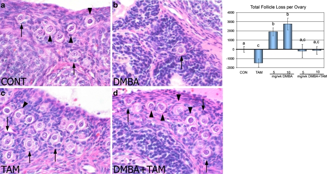
Introduction: we serendipitously observed a protective effect of tamoxifen against depletion of ovarian follicles by 7,12-dimethylbenzanthracene (DMBA), a chemical carcinogen, during a cancer prevention study. Such ovarian protection is being sought as an alternative approach to fertility preservation in human cancer patients.

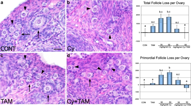
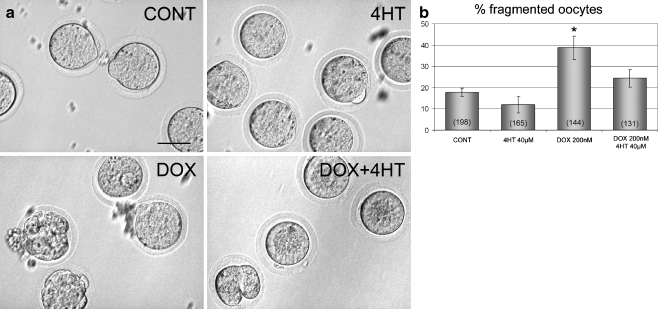
Methods: rats received tamoxifen (0, 1 mg or 2.5 mg/kg/d) and DMBA (0, 1, 2 mg/kg/wk) or cyclophosphamide (0, 35, 50 mg/kg/wk). Ovarian follicles were quantified and effects on fertility and litter size were tested. Cultured oocytes were exposed to chemotherapy drug doxorubicin, with or without 4-hydroxytamoxifen (4HT).

Results: DMBA and cyclophosphamide decreased the number of primordial and total follicles, and this reduction was prevented by tamoxifen. Cyclophosphamide tended to reduce fertility and lessened neonatal survival. Tamoxifen reversed these defects. Doxorubicin caused oocyte fragmentation which was prevented by 4HT.

Conclusions: tamoxifen decreases follicle loss and improves reproductive function following exposure to ovarian toxicants including chemotherapy drugs in the female rat.


Figures

Fig. 1
Fig. 1
H&E sections of the ovary from rats exposed to vehicle (CONT, a), DMBA (10 mg/wk, b), tamoxifen (TAM, c), or DMBA+TAM (d) for 2 weeks. Arrowheads = representative primordial follicles. Arrows = representative primary follicles. DMBA decreased follicular reserves in the ovary (b) compared with vehicle-treated controls (a) and the addition of tamoxifen inhibited this depletion (d). Tamoxifen alone elevated the number of follicles in the ovary compared with vehicle-treated controls (c). Numbers of follicle loss relative to vehicle-treated controls are shown for total (follicles at all developmental stages) follicle populations and are calculated by subtracting the control group mean value. The loss of follicles was expressed by a positive number whereas gain of follicles was expressed as a negative number. Different letters indicate significant differences among treatment groups
Fig. 2
Fig. 2
H&E sections of the ovary from rats exposed to vehicle (CONT, a), cyclophosphamide (Cy, 50 mg/kg, b), tamoxifen (TAM, c), or Cy+TAM (d) for 4 weeks. Arrowheads = representative primordial follicles. Arrows = representative primary follicles. Cy decreased total numbers of follicles (b) in the ovary compared with vehicle-treated controls (a) and the addition of tamoxifen inhibited this depletion (d). TAM did not affect the number of follicles in the ovary compared with vehicle-treated controls (c). Numbers of follicle loss relative to vehicle-treated controls are shown for total and primordial follicle populations. Cy treatment at 35 and 50 mg/kg/wk induced follicle loss compared with controls and the addition of TAM to both doses of Cy inhibited this depletion. Different letters indicate significant differences among treatment groups
Fig. 3
Fig. 3
a Mature oocytes exposed to vehicle, 4-hydroxytamoxifen (4HT), doxorubicin (DOX), or DOX+4HT for 24 h. Healthy oocytes surround by zona pellucida were observed after vehicle and 4HT treatment. Doxorubicin induces fragmentation in cultured oocytes, an effect that is antagonized by the addition of 4HT. b Quantitative analysis of the percentage of oocytes undergoing fragmentation. Oocytes incubated with doxorubicin showed increased fragmentation rate compared with vehicle-treated controls. This elevated fragmentation rate was inhibited by the addition of 4HT. The total number of oocytes cultured in each treatment group is indicated in parentheses inside the respective bar. * indicates a significant difference in comparison to vehicle-treated controls

References

    1. Absolom K, Eiser C, Turner L, Ledger W, Ross R, Davies H, Coleman R, Hancock B, Snowden J, Greenfield D. Ovarian failure following cancer treatment: current management and quality of life. Hum Reprod. 2008;23:2506–2512. - PubMed
    1. Simon B, Lee SJ, Partridge AH, Runowicz CD. Preserving fertility after cancer. CA Cancer J Clin. 2005;55:211–228. - PubMed
    1. Meirow D. Reproduction post-chemotherapy in young cancer patients. Mol Cell Endocrinol. 2000;169:123–131. - PubMed
    1. Pacey AA. Fertility issues in survivors from adolescent cancers. Cancer Treat Rev. 2007;33(7):646–655. - PubMed
    1. Lobo RA. Potential options for preservation of fertility in women. N Engl J Med. 2005;353(1):64–73. - PubMed

LinkOut - more resources