Review
. 2010;54(6-7):1151-63.
doi: 10.1387/ijdb.103106gs.

Hematopoietic stem cell emergence in the conceptus and the role of Runx1

Affiliations
Review

Hematopoietic stem cell emergence in the conceptus and the role of Runx1

Gemma Swiers et al. Int J Dev Biol. 2010.

Abstract

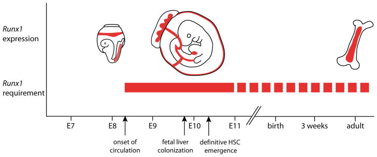
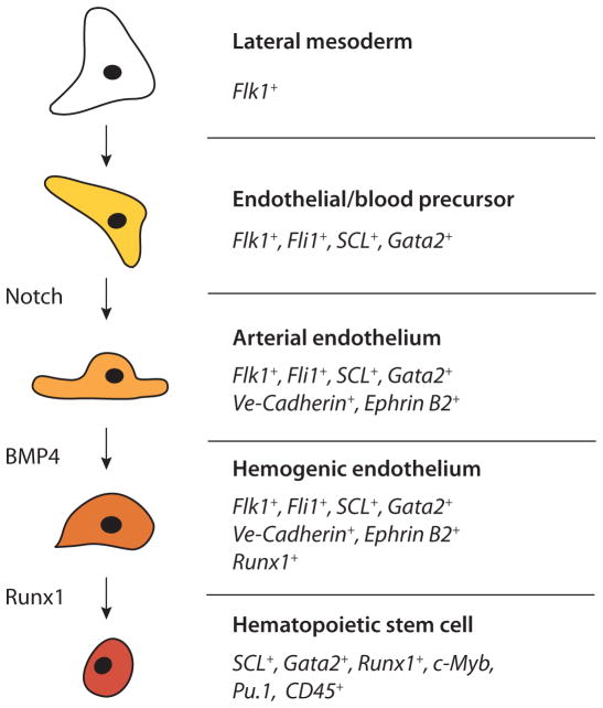
Hematopoietic stem cells (HSCs) are functionally defined as cells that upon transplantation into irradiated or otherwise immunocompromised adult organisms provide long-term reconstitution of the entire hematopoietic system. They emerge in the vertebrate conceptus around midgestation. Genetic studies have identified a number of transcription factors and signaling molecules that act at the onset of hematopoiesis, and have begun to delineate the molecular mechanisms underlying the formation of HSCs. One molecule that has been a particularly useful marker of this developmental event in multiple species is Runx1 (also known as AML1, Pebp2alpha). Runx1 is a sequence-specific DNA-binding protein, that along with its homologues Runx2 and Runx3 and their shared non-DNA binding subunit CBFbeta, constitute a small family of transcription factors called core-binding factors (CBFs). Runx1 is famous for its role in HSC emergence, and notorious for its involvement in leukemia, as chromosomal rearrangements and inactivating mutations in the human RUNX1 gene are some of the most common events in de novo and therapy-related acute myelogenous leukemia, myelodysplastic syndrome and acute lymphocytic leukemia. Here we will review the role of Runx1 in HSC emergence in the mouse conceptus and describe some of the genetic pathways that operate upstream and downstream of this gene. Where relevant, we will include data obtained from other species and embryonic stem (ES) cell differentiation cultures.


Figures

Fig. 1
Fig. 1. The outer boundaries of the proposed developmental window of the absolute Runx1 requirement in definitive blood cell generation (solid red bar)
A continued role for Runx1in specific hematopoietic cell types and lineages is represented by the dotted red bar. See text for additional explanation.
Fig. 2
Fig. 2. The Runx1 +23 enhancer recapitulates the hematopoietic specific expression pattern of Runx1
(A) Schematic of the Runx1 locus. Vertebrate Runx1 is transcribed from two promoters, the P1 and the P2. A 531 bp mouse-frog conserved enhancer was identified (Nottingham et al., 2007) and is located 23 kb downstream of the ATG in exon 1. (B) This +23 enhancer targets reporter gene expression to hematopoietic sites in the developing embryos, including all emerging HSCs. A transverse section through the dorsal aorta of an E10 transient transgenic embryo shows Xgal staining in emerging hematopoietic clusters (white arrowheads), in scattered cells of the endothelial wall (black arrowheads), and in a few mesenchymal cells (open arrowhead). Identical Xgal staining is seen in established mouse lines carrying the hsp68LacZ+23 transgene (not shown). (C) Targeted mutagenesis of putative transcription factor binding sites and chromatin IP (Nottingham et al., 2007), and trans-activation assays (Landry et al., 2008) placed the Runx1 +23 enhancer directly downstream of the ETS/GATA/SCL kernel (Liu et al., 2008; Pimanda et al., 2007b) that is active at the onset of developmental hematopoiesis.
Fig. 3
Fig. 3. Steps in hematopoietic stem cell formation, based on the model in which definitive blood is derived from so-called hemogenic endothelium
Arrows indicate the order of events only and should not be taken to represent direct transitions in all instances. Transcription factors, signaling pathways, and cell type specific genes active during this process are shown. This list is not exhaustive and is based on data available on mRNA and/or gene enhancer-mediated expression in mouse, chick, zebrafish and/or Xenopus embryos. In some instances there is a discrepancy in mRNA and protein expression. For example, VE-Cadherin protein is expressed on the cell membrane of mouse HSCs isolated from the AGM region, while in the chicken embryo aortic clusters do not express VE-Cadherin mRNA). This may be caused by the perdurance of protein following loss of mRNA synthesis. Pu.1 and c-Myb are expressed in pSp/AGM hematopoietic cells and play a role in fetal liver and adult bone marrow HSCs (Garcia et al., 2009; Iwasaki et al., 2005, Kim et al., 2004, Lieu and Reddy, 2009; Mukouyama et al., 1999; Sandberg et al., 2005). Thus, these transcription factors are likely to be expressed in AGM HSCs, although to our knowledge this was not formally shown.

References

    1. ADAMO L, NAVEIRAS O, WENZEL PL, MCKINNEY-FREEMAN S, MACK PJ, GRACIA-SANCHO J, SUCHY-DICEY A, YOSHIMOTO M, LENSCH MW, YODER MC, et al. Biomechanical forces promote embryonic haematopoiesis. Nature. 2009;459:1131–1135. - PMC - PubMed
    1. AIKAWA Y, NGUYEN LA, ISONO K, TAKAKURA N, TAGATA Y, SCHMITZ ML, KOSEKI H, KITABAYASHI I. Roles of HIPK1 and HIPK2 in AML1- and p300-dependent transcription, hematopoiesis and blood vessel formation. EMBO J. 2006;25:3955–3965. - PMC - PubMed
    1. ALVAREZ-SILVA M, BELO-DIABANGOUAYA P, SALAUN J, DIETERLEN-LIEVRE F. Mouse placenta is a major hematopoietic organ. Development. 2003;130:5437–5444. - PubMed
    1. ARAI F, HIRAO A, OHMURA M, SATO H, MATSUOKA S, TAKUBO K, ITO K, KOH GY, SUDA T. Tie2/angiopoietin-1 signaling regulates hematopoietic stem cell quiescence in the bone marrow niche. Cell. 2004;118:149–161. - PubMed
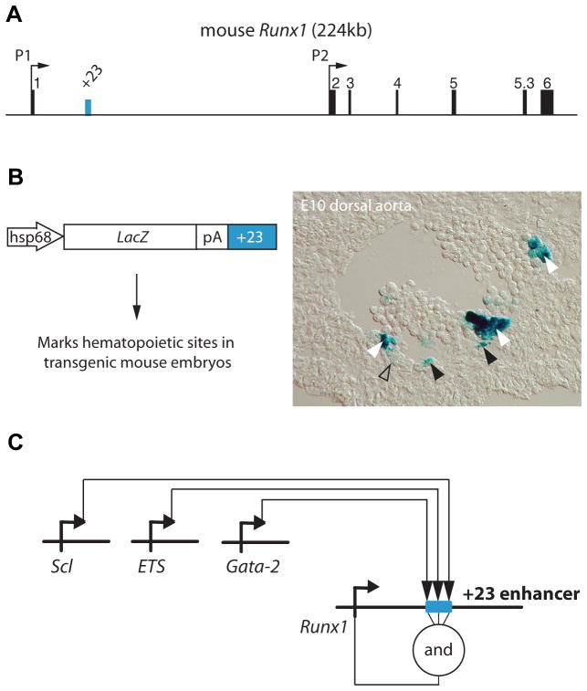
    1. BEE T, ASHLEY EL, BICKLEY SR, JARRATT A, LI PS, SLOANE-STANLEY J, GOTTGENS B, DE BRUIJN MF. The mouse Runx1 +23 hematopoietic stem cell enhancer confers hematopoietic specificity to both Runx1 promoters. Blood. 2009a;113:5121–5124. - PubMed

Publication types

MeSH terms

Substances