

High-fidelity hydrophilic probe for two-photon fluorescence lysosomal imaging
- PMID: 20712313
- PMCID: PMC2931774
- DOI: 10.1021/ja1057423
High-fidelity hydrophilic probe for two-photon fluorescence lysosomal imaging
Abstract
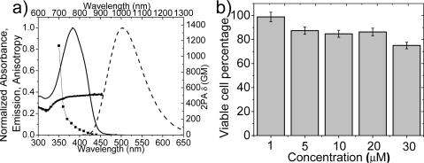
The synthesis and characterization of a novel two-photon-absorbing fluorene derivative, LT1, selective for the lysosomes of HCT 116 cancer cells, is reported. Linear and nonlinear photophysical and photochemical properties of the probe were investigated to evaluate the potential of the probe for two-photon fluorescence microscopy (2PFM) lysosomal imaging. The cytotoxicity of the probe was investigated to evaluate the potential of using this probe for live two-photon fluorescence biological imaging applications. Colocalization studies of the probe with commercial Lysotracker Red in HCT 116 cells demonstrated the specific localization of the probe in the lysosomes with an extremely high colocalization coefficient (0.96). A figure of merit was introduced to allow comparison between probes. LT1 has a number of properties that far exceed those of commercial lysotracker probes, including higher two-photon absorption cross sections, good fluorescence quantum yield, and, importantly, high photostability, all resulting in a superior figure of merit. 2PFM was used to demonstrate lysosomal tracking with LT1.
Figures


Similar articles
-
A series of fluorene-based two-photon absorbing molecules: synthesis, linear and nonlinear characterization, and bioimaging.J Org Chem. 2010 Jun 18;75(12):3975-82. doi: 10.1021/jo1005075. J Org Chem. 2010. PMID: 20481596 Free PMC article.
-
Amine-reactive fluorene probes: synthesis, optical characterization, bioconjugation, and two-photon fluorescence imaging.Bioconjug Chem. 2008 Dec;19(12):2559-67. doi: 10.1021/bc800415t. Bioconjug Chem. 2008. PMID: 19090700 Free PMC article.
-
Donor-acceptor-donor fluorene derivatives for two-photon fluorescence lysosomal imaging.J Org Chem. 2010 Jun 18;75(12):3965-74. doi: 10.1021/jo100554j. J Org Chem. 2010. PMID: 20481577 Free PMC article.
-
Two-photon STED spectral determination for a new V-shaped organic fluorescent probe with efficient two-photon absorption.Chemphyschem. 2011 Oct 24;12(15):2755-62. doi: 10.1002/cphc.201100456. Epub 2011 Aug 19. Chemphyschem. 2011. PMID: 21858908 Free PMC article.
-
Recent Advances in Two-Photon AIEgens and Their Application in Biological Systems.Chembiochem. 2021 Jun 2;22(11):1871-1883. doi: 10.1002/cbic.202000709. Epub 2021 Mar 3. Chembiochem. 2021. PMID: 33393721 Review.
Cited by
-
Integrin-targeting block copolymer probes for two-photon fluorescence bioimaging.Biomacromolecules. 2011 Feb 14;12(2):441-9. doi: 10.1021/bm1012212. Epub 2010 Dec 29. Biomacromolecules. 2011. PMID: 21190348 Free PMC article.
-
Molecular Fluorophores for Deep-Tissue Bioimaging.ACS Cent Sci. 2020 Aug 26;6(8):1302-1316. doi: 10.1021/acscentsci.0c00544. Epub 2020 Jul 23. ACS Cent Sci. 2020. PMID: 32875073 Free PMC article. Review.
-
Near-Infrared Bioimaging Using Two-photon Fluorescent Probes.Adv Healthc Mater. 2025 Jan;14(3):e2403272. doi: 10.1002/adhm.202403272. Epub 2024 Nov 21. Adv Healthc Mater. 2025. PMID: 39573885 Review.
-
Two-photon fluorescence microscopy imaging of cellular oxidative stress using profluorescent nitroxides.J Am Chem Soc. 2012 Mar 14;134(10):4721-30. doi: 10.1021/ja210315x. Epub 2012 Mar 1. J Am Chem Soc. 2012. PMID: 22380794 Free PMC article.
-
Synthesis of Near-Infrared Fluorescent Two-Photon-Absorbing Fluorenyl Benzothiadiazole and Benzoselenadiazole Derivatives.ACS Omega. 2016 Dec 7;1(6):1149-1156. doi: 10.1021/acsomega.6b00289. eCollection 2016 Dec 31. ACS Omega. 2016. PMID: 31457186 Free PMC article.
References
-
- Luzio J. P.; Pryor P. R.; Bright N. A. Nat. Rev. 2007, 8, 622–632. - PubMed
-
- Safig P.; Klumperman J. Nat. Rev. 2009, 10, 623–635. - PubMed
-
- Fehrenbacher N.; Jäättelä M. Cancer Res. 2005, 65, 2993–2995. - PubMed
-
- Glunde K.; Foss C. A.; Takagi T.; Wildes F.; Bhujwalla Z. Bioconjugate Chem. 2005, 16, 843–851. - PubMed
Publication types
MeSH terms
Substances
Grants and funding
LinkOut - more resources
Full Text Sources
