

doi: 10.1021/cb1001747.
Systematic analysis of helical protein interfaces reveals targets for synthetic inhibitors
Affiliations
- PMID: 20712375
- PMCID: PMC2955827
- DOI: 10.1021/cb1001747
Item in Clipboard
Systematic analysis of helical protein interfaces reveals targets for synthetic inhibitors
ACS Chem Biol.
.
Abstract
Synthetic inhibitors of protein-protein interactions are being discovered despite the inherent challenge in targeting large contact surfaces with small molecules. An analysis of available examples identifies common features of complexes that make them tractable for small molecules. We deduced that relative disposition and energetic contributions of "hot spot" residues provide a predictive scale for the potential of protein-protein interactions to be inhibited by small molecules. On the basis of this model, we analyzed the full set of helical protein interfaces in the Protein Data Bank to identify those that are potentially suitable candidates for synthetic ligands.
Figures
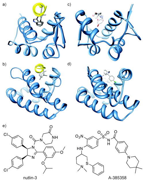
(a) The p53/HDM2 interaction (PDB code: 1YCR). A helix in the p53 activation domain resides in a deep hydrophobic groove. (b) The pro-apoptotic protein partner Bak bound to the anti-apoptotic protein Bcl-xL (PDB code: 1BXL). (c) Nutlin-3 binds to HDM2 in the same hydrophobic groove occupied by the p53 helix (PDB code: 1rv1). (d) ABT-785358 targets Bcl-xL at the site of its pro-apoptotic binding partners (PDB code: 2o22) (e) The structures of nutlin-3 and A-385358.
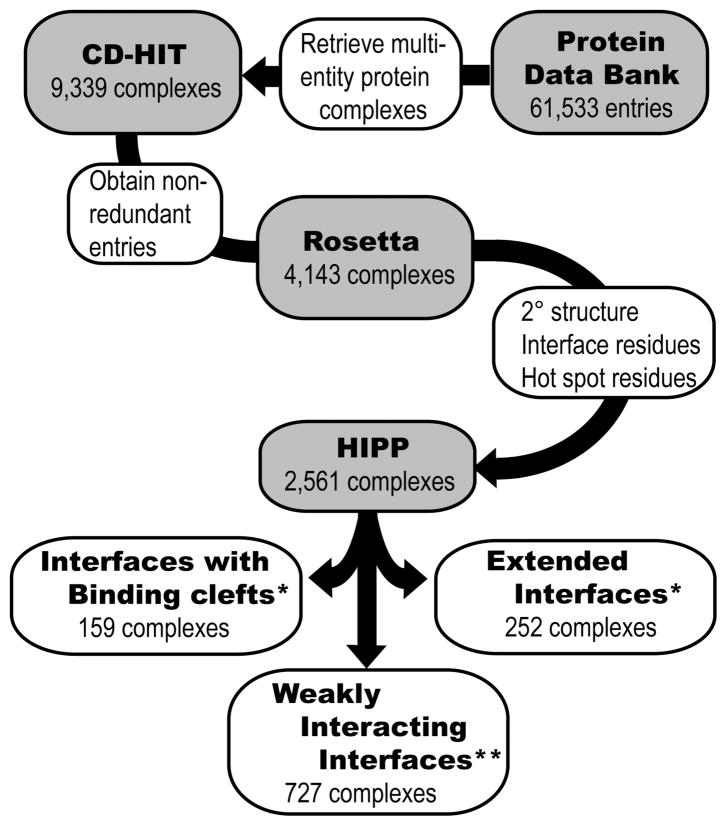
Evaluation of structures from the Protein Data Bank to identify and assess helical interfaces in protein–protein (HIPP) interactions. The helical interfaces were segregated based on binding interfaces and computational alanine scanning mutagenesis analysis. *ΔΔGavg ≥ 2 kcal/mol; ** ΔΔGavg = 1–2 kcal/mol.
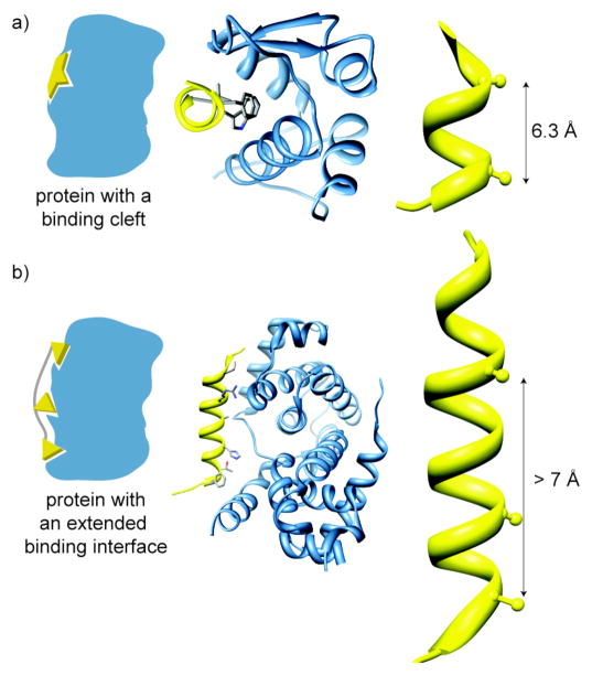
Helical Interfaces: we have divided helical protein-protein interactions between those that feature clefts for binding (a) and those with extended interfaces (b). The p53/MDM2 (PDB code: 1YCR) (a) and cyclin-dependent kinase6/D-type viral cyclin (PDB code: 1G3N) (b) complexes are representative examples of binding cleft and extended interfaces, respectively. The distance between flanking hot spot residues in the helix of the protein partner of a binding cleft target spans a radius of 7 Å or less (a) and greater than 7 Å but less than 30 Å for an extended interface target.
References
-
- Wells JA, McClendon CL. Reaching for high-hanging fruit in drug discovery at protein-protein interfaces. Nature. 2007;450:1001–1009. - PubMed
-
- Bogan AA, Thorn KS. Anatomy of hot spots in protein interfaces. J Mol Biol. 1998;280:1–9. - PubMed
-
- Moreira IS, Fernandes PA, Ramos MJ. Hot spots--a review of the protein-protein interface determinant amino-acid residues. Proteins. 2007;68:803–812. - PubMed
-
- Cunningham BC, Wells JA. High-resolution epitope mapping of hGH-receptor interactions by alanine-scanning mutagenesis. Science. 1989;244:1081–1085. - PubMed
-
- Kussie PH, Gorina S, Marechal V, Elenbaas B, Moreau J, Levine AJ, Pavletich NP. Structure of the MDM2 oncoprotein bound to the p53 tumor suppressor transactivation domain. Science. 1996;274:948–953. - PubMed
Publication types
MeSH terms
Substances
Grants and funding
LinkOut - more resources
Full Text Sources
Other Literature Sources
