. 2011 Mar 15;14(6):999-1010.
doi: 10.1089/ars.2010.3497. Epub 2010 Dec 7.

NOX3 NADPH oxidase couples transient receptor potential vanilloid 1 to signal transducer and activator of transcription 1-mediated inflammation and hearing loss

Affiliations

NOX3 NADPH oxidase couples transient receptor potential vanilloid 1 to signal transducer and activator of transcription 1-mediated inflammation and hearing loss

Debashree Mukherjea et al. Antioxid Redox Signal. .

Abstract

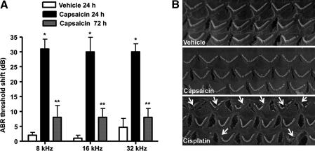
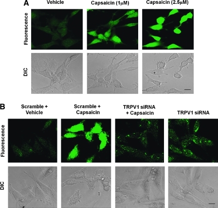
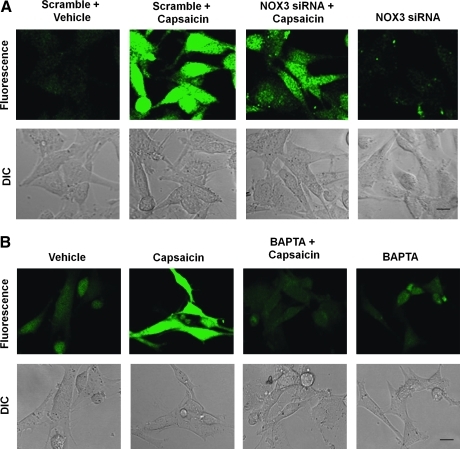
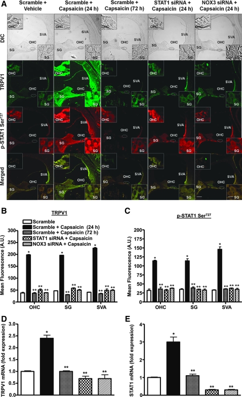
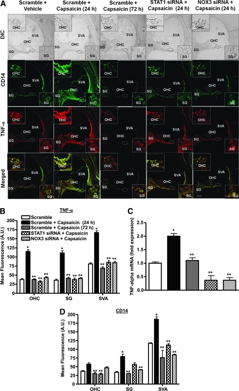
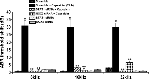
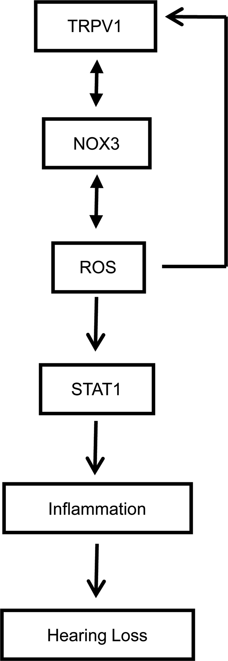
Transient receptor potential vanilloid 1 (TRPV1) is implicated in cisplatin ototoxicity. Activation of this channel by cisplatin increases reactive oxygen species generation, which contribute to loss of outer hair cells in the cochlea. Knockdown of TRPV1 by short interfering RNA protected against cisplatin ototoxicity. In this study, we examined the mechanism underlying TRPV1-mediated ototoxicity using cultured organ of Corti transformed cells (UB/OC-1) and rats. Trans-tympanic injections of capsaicin produced transient hearing loss within 24 h, which recovered by 72 h. In UB/OC-1 cells, capsaicin increased NOX3 NADPH oxidase activity and activation of signal transducer and activator of transcription 1 (STAT1). Intratympanic administration of capsaicin transiently increased STAT1 activity and expression of downstream proinflammatory molecules. Capsaicin produced a transient increase in CD14-positive inflammatory cells into the cochlea, which mimicked the temporal course of STAT1 activation but did not alter the expression of apoptotic genes or damage to outer hair cells. In addition, trans-tympanic administration of STAT1 short interfering RNA protected against capsaicin-induced hearing loss. These data suggest that activation of TRPV1 mediates temporary hearing loss by initiating an inflammatory process in the cochlea via activation of NOX3 and STAT1. Thus, these proteins represent reasonable targets for ameliorating hearing loss.


Figures

FIG. 1.
FIG. 1.
Transtympanic administration of capsaicin produces transient hearing loss. Capsaicin was administered by trans-tympanic injections to anesthetized male Wistar rats with the aid of a Zeiss operating microscope. The drug or vehicle was administered with 28G–30G needles in a volume of 50 μl. (A) ABR measurements performed 24 h later showed increased threshold shifts at all frequencies tested, but significant reductions from the 24 h levels at 72 h. *Statistically significant difference from vehicle-treated rats (p < 0.05, analysis of variance, n = 5–9 rats per group); **statistically significant difference from capsaicin (24 h)-treated group. (B) Scanning electron microscopy images of OHCs show no significant changes in morphology or loss 24 h after capsaicin administration. This is in contrast to cisplatin that produced significant degree of OHC damage or loss 72 h after intraperitoneal administration. Arrows indicate evidence of damage or loss of OHCs. Scanning electron microscopy images are a representative from one cochlea each from three different rats, with each showing similar changes. ABR, auditory brainstem response; OHC, outer hair cell.
FIG. 2.
FIG. 2.
Capsaicin increases ROS generation in UB/OC-1 cells. UB/OC-1 cells exposed to capsaicin demonstrated increased ROS generation as measured by H2DCFDA dye. (A) Cells were pretreated with different concentrations of capsaicin (0–2.5 μM) for 15 min, followed by incubation with H2DCFDA dye. The fluorescence intensity increased with increasing capsaicin concentrations. (B) Capsaicin-mediated increase in ROS generation was abrogated after knockdown of TRPV1 channels using siRNA. UB/OC-1 cells were transfected with TRPV1-siRNA for 48 h, and ROS generation was assessed in these cells after 15 min capsaicin treatment. TRPV1-siRNA alone did not alter the level of ROS generation. Data presented in (A) and (B) were replicated at least three times with similar results. Scale bar (lower right panels) represent 10 μm. H2DCFDA, 2′,7′-dichlorodihydrofluorescein diacetate; ROS, reactive oxygen species; siRNA, short interfering RNA; TRPV1, transient receptor potential 1. (For interpretation of the references to color in this figure legend, the reader is referred to the web version of this article at www.liebertonline.com/ars).
FIG. 3.
FIG. 3.
Capsaicin-induced ROS generation involves NOX3 activation. (A) UB/OC-1 cells exposed to capsaicin demonstrated increased ROS generation, as measured by H2DCFDA fluorescence. UB/OC-1 cells were pretreated with scramble or NOX3 siRNA for 48 h, the culture media were replaced with fresh media, and cultures were treated with vehicle or capsaicin (2.5 μM). Capsaicin increased ROS was abrogated by NOX3 siRNA. The transfection of cells with NOX3 siRNA alone did not alter H2DCFDA fluorescence. (B) Cells pretreated with vehicle or BAPTA-AM (10 μM) for 30 min before capsaicin administration showed reduced capsaicin-mediated ROS generation. The addition of BAPTA-AM alone was not associated with any appreciable change in ROS generation. Scale bar (lower right panel) represents 10 μm. Data presented in (A) and (B) were replicated at least three times with similar results. BAPTA-AM, 1,2-Bis(2-aminophenoxy)ethane-N,N,N′,N′-tetraacetic acid tetrakis (acetoxymethyl ester). (For interpretation of the references to color in this figure legend, the reader is referred to the web version of this article at www.liebertonline.com/ars).
FIG. 4.
FIG. 4.
Capsaicin increases STAT1 activity in UB/OC-1 cells and in the rat cochlea. (A) UB/OC-1 cells were incubated with vehicle (0) or capsaicin (2.5 μM) for up to 2 h and nuclear lysates were prepared from each time point and used for determination of Ser727 p-STAT1 and total STAT1 by Western blotting. Capsaicin increased Ser727 p-STAT1 levels over total STAT1, indicative of STAT1 activation. *Statistically significant difference from vehicle-treated cells (p < 0.05; n = 4). (B) Capsaicin increased Ser727 p-STAT1 in the rat cochlea. Rats were anesthetized and then administered capsaicin by trans-tympanic injections. Cochleae were isolated 24 or 72 h later, perfused with 4% glutaraldehyde, decalcified, and paraffin imbedded, and the resulting sections used for Ser727 p-STAT1 immunocytochemistry. Capsaicin increased Ser727 p-STAT1 levels throughout the cochlea, but especially in the SVA, OHCs, and SG cells (see insets). (C) Similar increases in total STAT1 were observed in adjacent cochlear sections. Data presented in (B) and (C) are representative sections from three different rats (one cochlea each) per group. Insets show enlarged sections of OHCs and SG. Scale bars (right lower panel) represent 50 and 10 μm (for insets). SG, spiral ganglion; STAT1, signal transducer and activator of transcription 1; SVA, stria vascularis. (For interpretation of the references to color in this figure legend, the reader is referred to the web version of this article at www.liebertonline.com/ars).
FIG. 5.
FIG. 5.
Capsaicin-mediated STAT1 activation in vivo is transient and involves ROS generation. (A) Rats were anesthetized and administered scrambled siRNA (scramble), STAT1, or NOX3 siRNAs by trans-tympanic injections. Two days later capsaicin was administered by intratympanic injections for 24 or 72 h. Cochleae were isolated, sectioned, and used for p-STAT1 Ser727 and TRPV1 immunolabeling studies. Capsaicin increased both TRPV1 (green) and p-STAT1 Ser727 (red) immunoreactivity at 24 h in SVA, OHC, and SG. However, these increases recovered to baseline levels by 72 h. Animals pretreated with STAT1 or NOX3 siRNA did not show any induction of p-STAT1 Ser727 and TRPV1 immunolabeling. Merged images (yellow) show colocalization of TRPV1 and p-STAT1 Ser727, suggesting possible functional interactions between these two proteins. Images shown are representatives of cochleae from four different animals showing similar results. Insets show enlarged sections of OHCs and SGs. Scale bars (lower right panel) represent 50 and 10 μm for insets. (B, C) Quantification of TRPV1 and p-STAT1 Ser727 immunolabeling indicate significant increases in the levels at the 24 h period after capsaicin administration but substantially reduced levels of these proteins were evident at 72 h or in STAT1 or NOX3 siRNAs pretreated groups. (D, E) Quantification of mRNA from rat cochleae by real-time polymerase chain reaction indicated significant increases in STAT1 and TRPV1 expression at 24 h but recovery to essentially baseline levels at 72 h. Knockdown of STAT1 or NOX3 by siRNAs abolished the increases in these transcripts. *Statistically significant difference from scramble siRNA-treated rats; **statistically significant difference from scramble + capsaicin-24 h treated rats (p < 0.05; n = 4). (For interpretation of the references to color in this figure legend, the reader is referred to the web version of this article at www.liebertonline.com/ars).
FIG. 6.
FIG. 6.
Capsaicin increases expression of STAT1-targeted genes in the cochlea. Rats were administered scrambled (scramble), STAT1, or NOX3 siRNAs via the trans-tympanic route, followed by vehicle or capsaicin. Cochleae were retrieved 24 or 72 h later, processed for coimmunolabeling for CD14 (green) and TNF-α (red) and RNA preparation. (A) In control animals (scramble), CD14 and TNF-α immunoreactivity was localized to the SVA and less to the OHC and SG cells. These proteins showed colocalization in cochlear merged images (yellow). Capsaicin produced substantial increases in labeling of these two proteins in these regions at 24 h but not at 72 h. However, pretreatment with STAT1 and NOX3 siRNAs abolished the regional increases in these proteins. Both STAT1 and NOX3 siRNAs reduced capsaicin-stimulated CD14 and TNF-α immunolabeling. Insets are enlarged sections of OHCs and SGs. Scale bars (right lower panels) represent 50 and 10 μm for insets. Images shown are representatives from cochleae from four rats showing similar results. (B, D) Quantification of immunofluorescence depicted in (A). (C) TNF-α mRNA obtained from the groups of rats treated similarly as in (A). *Statistically significant difference from scramble + vehicle-treated rats; **statistically significant difference from scramble + capsaicin-24 h treated rats (p < 0.05; n = 4). TNF-α, tumor necrosis factor-α. (For interpretation of the references to color in this figure legend, the reader is referred to the web version of this article at www.liebertonline.com/ars).
FIG. 7.
FIG. 7.
Trans-tympanic delivery of STAT1 siRNA protects against hearing loss. Rats were anesthetized and then administered scrambled (scramble) STAT1 or NOX3 siRNAs. Two days later, these animals were administered vehicle or capsaicin and tested 24 h later. Capsaicin produced a ∼30 dB threshold shift at all frequencies examined, which was attenuated by trans-tympanic administration of STAT1 or NOX3 siRNAs. *Statistically significant difference from animals administered a scramble siRNA sequence; **statistically significant reduction in the ABR threshold shifts from capsaicin-24 h treated group (p < 0.05; n = 6).
FIG. 8.
FIG. 8.
Model of capsaicin-induced inflammation in the cochlea. Capsaicin activates TRPV1, which allows entry of Ca2+ into the cell and increased ROS production via NOX3 activation of STAT1. ROS also produce positive feedback regulation of NOX3 and TRPV1 expression. STAT1 promotes induction of a number of proinflammatory genes that contribute to cochlear inflammation and hearing loss.

Similar articles

Cited by

References

    1. Alawi K. Keeble J. The paradoxical role of the transient receptor potential vanilloid 1 receptor in inflammation. Pharmacol Ther. 2010;125:181–195. - PubMed
    1. Bánfi B. Malgrange B. Knisz J. Steger K. Dubois-Dauphin M. Krause K-H. NOX3, a superoxide-generating NADPH oxidase of the inner ear. J Biol Chem. 2004;279:46065–46072. - PubMed
    1. Bielefeld EC. Hu BH. Harris KC. Henderson D. Damage and threshold shift resulting from cochlear exposure to paraquat-generated superoxide. Hear Res. 2005;207:35–42. - PMC - PubMed
    1. Birder LA. Nakamura Y. Kiss S. Nealen ML. Barrick S. Kanai AJ. Wang E. Ruiz G. De Groat WC. Apodaca G. Watkins S. Caterina MJ. Altered urinary bladder function in mice lacking the vanilloid receptor TRPV1. Nat Neurosci. 2002;5:856–860. - PubMed
    1. Christoph T. Grünweller A. Mika J. Schäfer MK. Wade EJ. Weihe E. Erdmann VA. Frank R. Gillen C. Kurreck J. Silencing of vanilloid receptor TRPV1 by RNAi reduces neuropathic and visceral pain in vivo. Biochem Biophys Res Commun. 2006;350:238–243. - PubMed

Publication types

MeSH terms