

Confusion after spine injury: cerebral fat embolism after traumatic rupture of a Tarlov cyst: case report
- PMID: 20712856
- PMCID: PMC2930635
- DOI: 10.1186/1471-227X-10-18
Confusion after spine injury: cerebral fat embolism after traumatic rupture of a Tarlov cyst: case report
Abstract
Background: Acute low back pain is a very common symptom and reason for many medical consultations. In some unusual circumstances it could be linked to a rare aetiology.
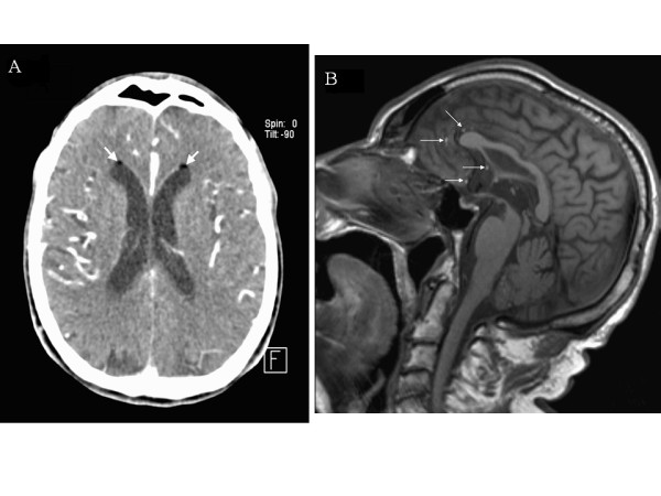
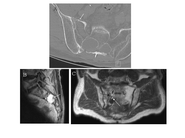
Case presentation: We report a 70-year-old man with an 8-month history of left posterior thigh and leg pain who had sudden confusion after a fall from standing. It was due to cerebral fat embolism suspected by computed tomography scan, later confirmed by brain magnetic resonance imaging (MRI). A spinal MRI scan was then performed and revealed a sacral fracture which drained into an unknown perineurial cyst (Tarlov cyst). Under medical observation the patient fully recovered within three weeks.
Conclusions: Sacral perineurial cysts are rare, however they remain a potential cause of lumbosacral radiculopathy.
Figures
Similar articles
-
Tarlov cyst as a rare cause of S1 radiculopathy: A case report.Arch Phys Med Rehabil. 2001 May;82(5):689-90. doi: 10.1053/apmr.2001.22353. Arch Phys Med Rehabil. 2001. PMID: 11346849
-
An unusual case of CSF leak following post-traumatic rupture of a sacral meningeal cyst.Cephalalgia. 2015 Oct;35(12):1130-2. doi: 10.1177/0333102414566202. Epub 2015 Jan 8. Cephalalgia. 2015. PMID: 25573898 No abstract available.
-
Symptomatic sacral perineurial (Tarlov) cysts.Coll Antropol. 2009 Dec;33(4):1401-3. Coll Antropol. 2009. PMID: 20102100
-
Diagnosis and management of sacral Tarlov cysts. Case report and review of the literature.Neurosurg Focus. 2003 Aug 15;15(2):E15. doi: 10.3171/foc.2003.15.2.15. Neurosurg Focus. 2003. PMID: 15350046 Review.
-
Sacral fracture associated with a Tarlov cyst causing an anterior sacral CSF fistula and intraventricular fat emboli - a case report and review of the literature.Br J Neurosurg. 2024 Jun;38(3):591-595. doi: 10.1080/02688697.2021.1940848. Epub 2021 Aug 16. Br J Neurosurg. 2024. PMID: 34397315 Review.
Cited by
-
Comparative outcomes of the two types of sacral extradural spinal meningeal cysts using different operation methods: a prospective clinical study.PLoS One. 2013 Dec 26;8(12):e83964. doi: 10.1371/journal.pone.0083964. eCollection 2013. PLoS One. 2013. PMID: 24386317 Free PMC article. Clinical Trial.
-
Low pressure headache and cerebral fat embolism from a sacral fracture through a Tarlov cyst: a case report.J Med Case Rep. 2023 Oct 7;17(1):444. doi: 10.1186/s13256-023-04142-2. J Med Case Rep. 2023. PMID: 37803426 Free PMC article.
-
Rare clinical presentations of perineural cysts besides radicular pain.Korean J Pain. 2012 Oct;25(4):283-4. doi: 10.3344/kjp.2012.25.4.283. Epub 2012 Oct 4. Korean J Pain. 2012. PMID: 23091694 Free PMC article. No abstract available.
-
Management of Tarlov cysts: an uncommon but potentially serious spinal column disease-review of the literature and experience with over 1000 referrals.Neuroradiology. 2024 Jan;66(1):1-30. doi: 10.1007/s00234-023-03226-6. Epub 2023 Oct 13. Neuroradiology. 2024. PMID: 37828278 Free PMC article. Review.
-
Large presacral Tarlov cysts in pregnancy.Clin Case Rep. 2022 May 12;10(5):e05837. doi: 10.1002/ccr3.5837. eCollection 2022 May. Clin Case Rep. 2022. PMID: 35592042 Free PMC article.
References
-
- Tarlov IM. Perineural cysts of the spinal nerve roots. Arch Neurol Psychiatry. 1938;40:1067–1074.
-
- Tarlov IM. Cysts, perineurial, of the sacral roots: another cause, removable, of sciatic pain. JAMA. 1948;138:740–744. - PubMed
Publication types
MeSH terms
LinkOut - more resources
Full Text Sources
Medical
