. 2010 Aug 16:8:98.
doi: 10.1186/1477-7827-8-98.

Whole ovary immunohistochemistry for monitoring cell proliferation and ovulatory wound repair in the mouse

Affiliations

Whole ovary immunohistochemistry for monitoring cell proliferation and ovulatory wound repair in the mouse

Rajasekhar Singavarapu et al. Reprod Biol Endocrinol. .

Abstract

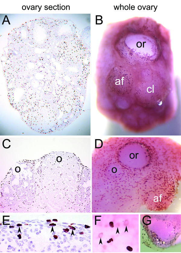
Background: Ovarian surface epithelial cells are thought to be a precursor cell type for ovarian carcinoma. It has been proposed that an increased rate of ovarian surface epithelial cell proliferation during ovulatory wound repair contributes to the accumulation of genetic changes and cell transformation. The proliferation of ovarian surface epithelial cells during ovulatory wound repair has been studied primarily using immunohistochemical staining of paraffin-embedded ovary sections. However, such analyses require complex reconstruction from serially-cut ovary sections for the visualization and quantification of the cells on the ovarian surface. In order to directly visualize the proliferation and organization of the ovarian surface epithelial cells, we developed a technique for immunohistochemical staining of whole mouse ovaries. Using this method, we analyzed cell proliferation and morphologic changes in mouse ovarian surface epithelial cells during follicle growth and ovulatory wound repair.

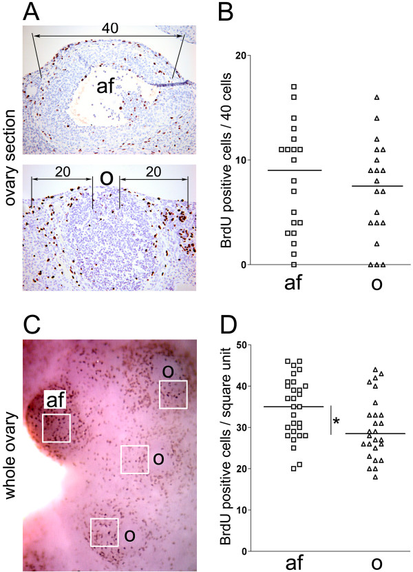
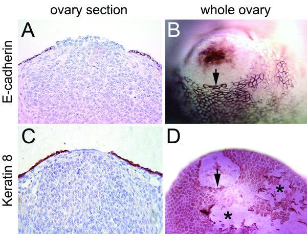
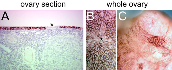
Methods: Three-week old FVB/N female mice were superovulated by sequential administration of pregnant mare's serum gonadotropin (PMSG) and human chorionic gonadotropin (hCG). Ten hours after hCG administration, mice were given 5-bromo-2-deoxyuridine (BrdU) and euthanized two hours after BrdU administration for ovary isolation. The levels of incorporated BrdU in the ovarian surface epithelial cells were measured by staining paraffin-embedded ovary sections and whole ovaries with the BrdU antibody. Re-epithelialization of the ovarian surface after ovulatory rupture was visualized by immunohistochemical staining with E-cadherin and Keratin 8 in paraffin-embedded ovary sections and whole ovaries.

Results: We determined that active proliferation of ovarian epithelial surface cells primarily occurs during antral follicle formation and, to a lesser extent, in response to an ovulatory wound. We also demonstrated that ovarian surface epithelial cells exhibit a circular organization around the wound site

Conclusion: Whole ovary immunohistochemistry enables efficient and comprehensive three-dimensional visualization of ovarian surface epithelial cells without the need for laborious reconstruction from immunohistochemically-stained serial ovary sections.


Figures

Figure 1
Figure 1
BrdU staining of ovary sections and whole ovaries in superovulated mice. BrdU was administered to the mice 10 hours after hCG injection and the ovaries were isolated 2 hours after BrdU injection. The distribution of cells with incorporated BrdU was determined by staining paraffin-embedded ovary sections (A, C, E) and whole ovaries (B, D, F, G) with anti-BrdU antibody. Arrowheads indicate BrdU positive cells below the ovarian surface epithelial layer. Abbreviations: af, antral follicle; cl, corpus luteum; o, ovulatory wound; or, recent ovulatory wound.
Figure 2
Figure 2
Quantification of BrdU incorporation during antral follicle formation and ovulatory wound repair in ovary sections (A, B) and whole ovaries (C, D). (A) In ovary sections, the number of BrdU positive cells associated with the antral follicles (af) was determined by scoring forty cells covering the surface of each antral follicle. The number of BrdU positive cells associated with corpora lutea containing healing ovulatory wounds (o) was determined by scoring twenty cells covering each side of the ovulatory wound. (B) Dot plot of the number of BrdU positive cells associated with antral follicles and ovulatory wounds. Each rectangle represents one antral follicle (N = 20) and each triangle represents one corpus luteum with an ovulatory wound (N = 20). The horizontal bars represent the mean values. P = 0.5026. (C) In the whole ovary, the number of BrdU positive cells associated with antral follicles (af) and ovulatory wounds (o) was determined by counting positive cells per square unit. (D) Dot plot of the number of BrdU positive cells associated with antral follicles (af) and ovulatory wounds (o). Each rectangle represents one antral follicle (N = 29) and each triangle represents one corpus luteum with an ovulatory wound (N = 26). The horizontal bars represent the mean values. *P = 0.0175.
Figure 3
Figure 3
Re-epithelialization of the ovarian surface after ovulatory rupture. Ovarian surface epithelial cells were visualized by immunohistochemical staining with E-cadherin (A, B) and Keratin 8 (C, D) in paraffin-embedded ovary sections (A, C) and whole ovaries (B, D). The arrows point to well-organized epithelial edges around the ovulatory wound. The asterisks indicate disorganized epithelia where the ovarian surface was artificially scraped by ovary manipulation.
Figure 4
Figure 4
E-cadherin expression in the ovarian surface epithelial cells. Immunohistochemical staining for E-cadherin in a paraffin-embedded ovary section (A) and whole ovaries (B, C). The asterisks indicate groups of epithelial cells in which E-cadherin is expressed at a low level. Distinct patches of ovarian surface epithelial cells with variable levels of E-cadherin expression are detected by whole ovary immunohistochemistry (C).

References

    1. Bernal A, Mendez-Moran L, Fajardo-Gutierrez A, Gonzalez-Lira G, Escudero P, Ortiz H. Univariate and multivariate analysis of risk factors for ovarian cancer: case-control study, Mexico City. Arch Med Res. 1995;26:245–249. - PubMed
    1. Perez RP, Godwin AK, Hamilton TC, Ozols RF. Ovarian cancer biology. Semin Oncol. 1991;18:186–204. - PubMed
    1. Purdie DM, Bain CJ, Siskind V, Webb PM, Green AC. Ovulation and risk of epithelial ovarian cancer. Int J Cancer. 2003;104:228–232. doi: 10.1002/ijc.10927. - DOI - PubMed
    1. Auersperg N, Edelson MI, Mok SC, Johnson SW, Hamilton TC. The biology of ovarian cancer. Semin Oncol. 1998;25:281–304. - PubMed
    1. Murdoch WJ, Townsend RS, McDonnel AC. Ovulation-induced DNA damage in ovarian surface epithelial cells of ewes: prospective regulatory mechanisms of repair/survival and apoptosis. Biol Reprod. 2001;65:1417–1424. doi: 10.1095/biolreprod65.5.1417. - DOI - PubMed

Publication types

MeSH terms