

p53 nuclear accumulation and ERalpha expression in ductal hyperplasia of breast in a cohort of 215 Chinese women
- PMID: 20712900
- PMCID: PMC2936335
- DOI: 10.1186/1756-9966-29-112
p53 nuclear accumulation and ERalpha expression in ductal hyperplasia of breast in a cohort of 215 Chinese women
Abstract
Introduction: Women with ductal hyperplasia including usual ductal hyperplasia (UDH) and atypical ductal hyperplasia (ADH) have an increased risk of developing invasive ductal carcinoma (IDC) of breast. The importance of several molecular markers in breast cancer has been of considerable interest during recent years such as p53 and estrogen receptor alpha (ERalpha). However, p53 nuclear accumulation and ERalpha expression have not been assessed in ductal hyperplasia co-existing with ductal carcinoma in situ (DCIS) or IDC versus pure ductal hyperplasia without DCIS or IDC.
Materials and methods: We investigated p53 nuclear accumulation and ERalpha expression in breast ductal hyperplasia in a cohort of 215 Chinese women by immunohistochemistry (IHC), which included 129 cases of pure ductal hyperplasia, 86 cases of ductal hyperplasia co-existing with DCIS (41 cases) or IDC (45 cases).
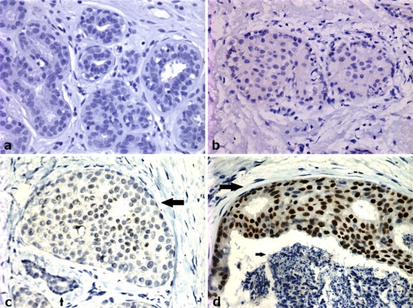
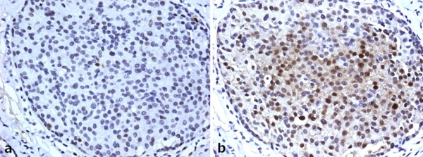
Results: Nuclear p53 accumulation was identified in 22.8% of ADH (31/136), 41.5% of DCIS (17/41) and 42.2% of IDC (19/45), and no case of UDH (0/79). No difference in nuclear p53 accumulation was observed between pure ADH and ADH co-existing with DCIS (ADH/DCIS) or IDC (ADH/IDC) (P>0.05). The positive rate of ERalpha expression was lower in ADH (118/136, 86.8%) than that in UDH (79/79, 100%) (P<0.001), but higher than that in DCIS (28/41, 68.3%) or IDC (26/45, 57.8%) respectively (P<0.001). The frequency of ERalpha expression was lower in ADH/DCIS (23/29, 79.31%) and ADH/IDC (23/30, 76.67%) than that in pure ADH (72/77, 93.51%) respectively (P<0.05). There was a negative weak correlation between p53 nuclear accumulation and ERalpha expression as for ADH (coefficient correlation -0.51; P<0.001).
Conclusions: Different pathological types of ductal hyperplasia of breast are accompanied by diversity in patterns of nuclear p53 accumulation and ERalpha expression. At least some pure ADH is molecularly distinct from ADH/CIS or ADH/IDC which indicated the two types of ADH are molecularly distinct entities although they have the same morphological appearance.
Figures

Similar articles
-
14-3-3ζ promoted invasion and lymph node metastasis of breast invasive ductal carcinoma with HER2 overexpression.Pathol Res Pract. 2021 Nov;227:153619. doi: 10.1016/j.prp.2021.153619. Epub 2021 Sep 22. Pathol Res Pract. 2021. PMID: 34560418
-
Expression pattern and methylation of estrogen receptor α in breast intraductal proliferative lesions.Oncol Rep. 2016 Oct;36(4):1868-74. doi: 10.3892/or.2016.4988. Epub 2016 Aug 1. Oncol Rep. 2016. PMID: 27498697 Free PMC article.
-
Evidence of chromosomal alterations in pure usual ductal hyperplasia as a breast carcinoma precursor.Oncol Rep. 2008 Jun;19(6):1469-75. Oncol Rep. 2008. PMID: 18497952
-
HER2 as a prognostic factor in breast cancer.Oncology. 2001;61 Suppl 2:67-72. doi: 10.1159/000055404. Oncology. 2001. PMID: 11694790 Review.
-
The diagnosis and management of pre-invasive breast disease: ductal carcinoma in situ (DCIS) and atypical ductal hyperplasia (ADH)--current definitions and classification.Breast Cancer Res. 2003;5(5):254-7. doi: 10.1186/bcr623. Epub 2003 Jul 29. Breast Cancer Res. 2003. PMID: 12927035 Free PMC article. Review.
Cited by
-
Estrogen receptor-α promoter methylation is a biomarker for outcome prediction of cisplatin resistance in triple-negative breast cancer.Oncol Lett. 2018 Mar;15(3):2855-2862. doi: 10.3892/ol.2017.7637. Epub 2017 Dec 19. Oncol Lett. 2018. PMID: 29456719 Free PMC article.
-
Promoter methylation status and expression of estrogen receptor alpha in familial breast cancer patients.Tumour Biol. 2012 Apr;33(2):413-20. doi: 10.1007/s13277-011-0234-x. Epub 2011 Sep 16. Tumour Biol. 2012. PMID: 21922275
-
Estrogen receptor-alpha promoter methylation in sporadic basal-like breast cancer of Chinese women.Tumour Biol. 2011 Aug;32(4):713-9. doi: 10.1007/s13277-011-0172-7. Epub 2011 Apr 6. Tumour Biol. 2011. PMID: 21625942
-
CXCL12-CXCR4 axis promotes the natural selection of breast cancer cell metastasis.Tumour Biol. 2014 Aug;35(8):7765-73. doi: 10.1007/s13277-014-1816-1. Epub 2014 May 9. Tumour Biol. 2014. PMID: 24810923 Free PMC article.
-
Analysis of miR-205 and miR-155 expression in the blood of breast cancer patients.Chin J Cancer Res. 2013 Feb;25(1):46-54. doi: 10.3978/j.issn.1000-9604.2012.11.04. Chin J Cancer Res. 2013. PMID: 23372341 Free PMC article.
References
-
- Boyle P, Levin B. World Cancer Report. International Agency for Research on Cancer. 2003.
-
- Steinman S, Wang J, Bourne P, Yang Q, Tang P. Expression of cytokeratin markers, ER-alpha, PR, HER-2/neu, and EGFR in pure ductal carcinoma in situ (DCIS) and DCIS with co-existing invasive ductal carcinoma (IDC) of the breast. Ann Clin Lab Sci. 2007;37:127–134. - PubMed
-
- Arpino G, Laucirica R, Elledge RM. Premalignant and in situ breast disease: biology and clinical implications. Ann Intern Med. 2005;143:446–457. - PubMed
Publication types
MeSH terms
Substances
LinkOut - more resources
Full Text Sources
Medical
Research Materials
Miscellaneous
