

Regional calcineurin subunit B isoform expression in rat hippocampus following a traumatic brain injury
- PMID: 20713027
- PMCID: PMC2949526
- DOI: 10.1016/j.brainres.2010.08.029
Regional calcineurin subunit B isoform expression in rat hippocampus following a traumatic brain injury
Abstract
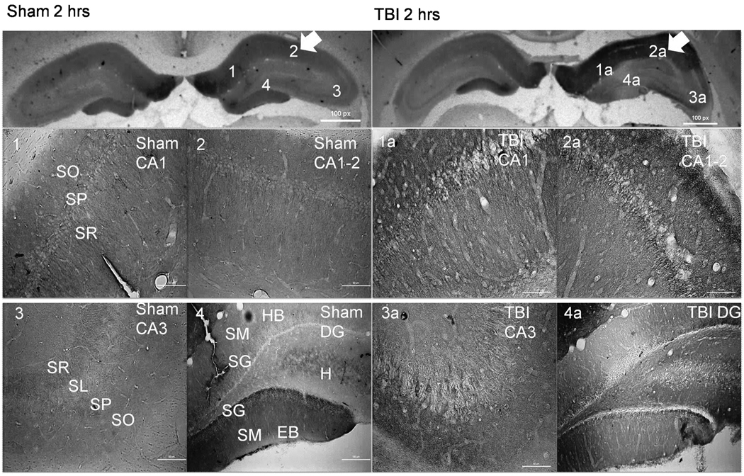
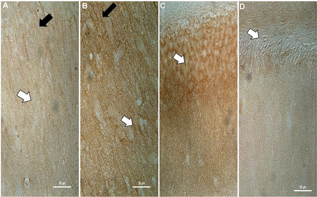
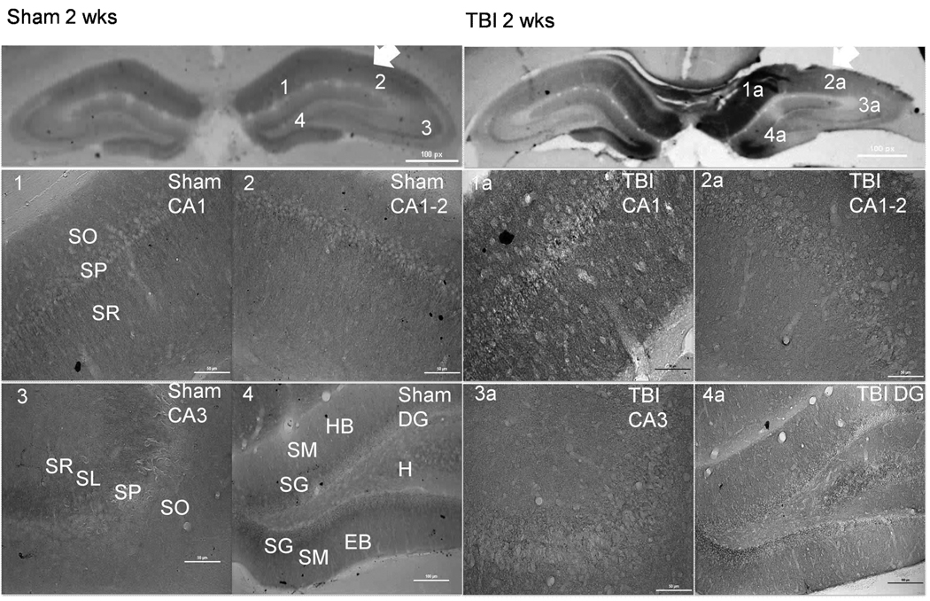
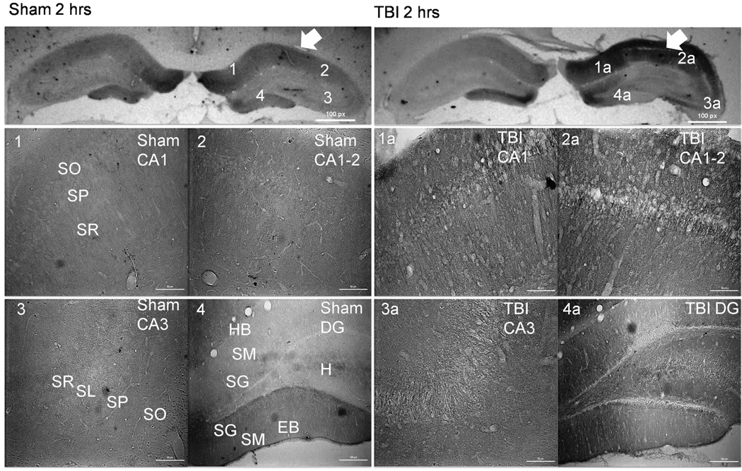
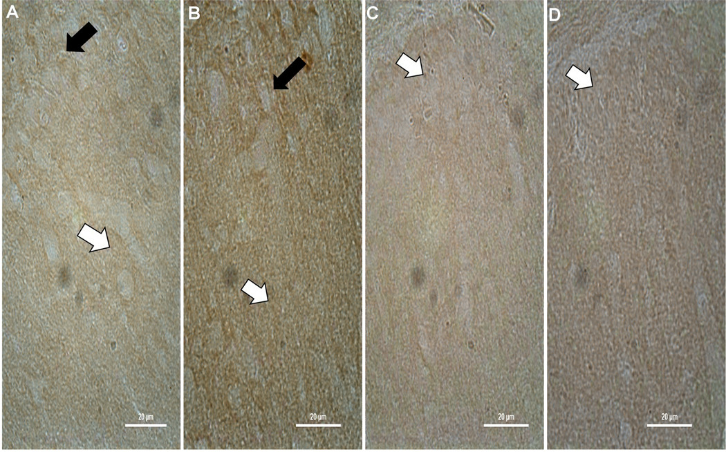
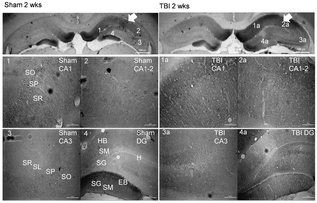
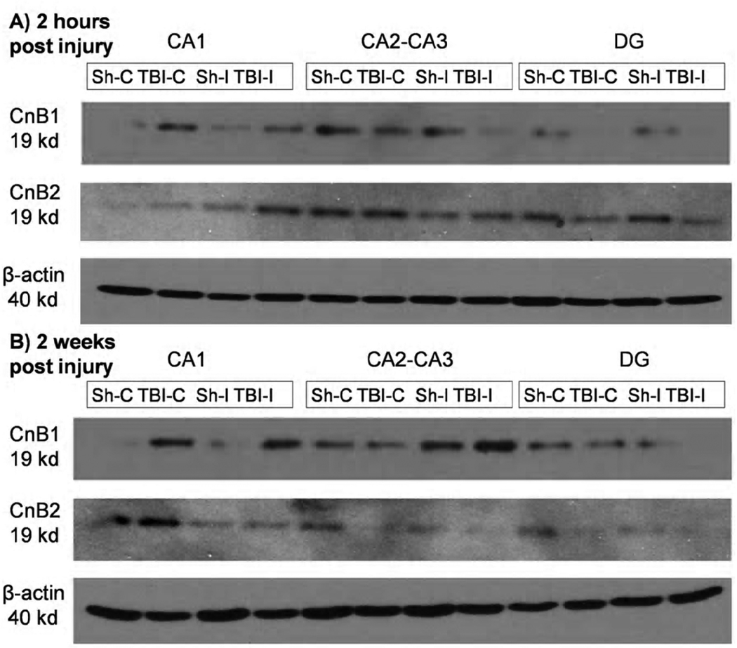
Calcineurin subunit isoforms are implicated in long term potentiation, long term depression, and structural plasticity. Calcineurin inhibitors benefit axonal damage, cellular dysfunction, and cognitive outcomes in animal models of traumatic brain injury (TBI). Distribution of the catalytic calcineurin A subunit is altered and calcineurin activity increased following fluid percussion injury. Alterations in calcineurin subunit A isoform distribution within the hippocampus also occur post controlled cortical impact (CCI) demonstrating a reduction in catalytic subunit distribution in CA1-2 dendritic fields. Furthermore the effect of TBI on the regulatory subunit, calcineurin B, is unknown. Understanding the role of both subunits is necessary to effectively target alterations in calcineurin signaling as current calcineurin inhibitors, such as cyclosporin A and FK-506, rely upon binding sites on both subunits for complete inhibition. The effect of moderate CCI on the expression and distribution of calcineurin B isoforms within the hippocampus was examined at 2h and 2weeks post injury. Calcineurin B isoforms showed increased expression throughout the CA1 and CA2 while there was a decrease in expression within the ipsilateral dentate gyrus. Alterations in CnB isoform expression within the CA1, CA1-2, and dentate gyrus have significant implications for persistent hippocampal dysfunction following TBI. Regional changes in regulatory subunit expression may alter the effect of calcineurin inhibitors regionally following a traumatic brain injury.
Published by Elsevier B.V.
Figures
Similar articles
-
Expression of protein phosphatase 2B (calcineurin) subunit A isoforms in rat hippocampus after traumatic brain injury.J Neurotrauma. 2010 Jan;27(1):109-20. doi: 10.1089/neu.2009.1072. J Neurotrauma. 2010. PMID: 19751097 Free PMC article.
-
Blockade of Astrocytic Calcineurin/NFAT Signaling Helps to Normalize Hippocampal Synaptic Function and Plasticity in a Rat Model of Traumatic Brain Injury.J Neurosci. 2016 Feb 3;36(5):1502-15. doi: 10.1523/JNEUROSCI.1930-15.2016. J Neurosci. 2016. PMID: 26843634 Free PMC article.
-
Alteration in the immunoreactivity of the calcineurin subunits after ischemic hippocampal damage.Neuroscience. 1992 Aug;49(3):545-56. doi: 10.1016/0306-4522(92)90225-q. Neuroscience. 1992. PMID: 1323805
-
A significant increase in both basal and maximal calcineurin activity following fluid percussion injury in the rat.J Neurotrauma. 2005 Apr;22(4):476-90. doi: 10.1089/neu.2005.22.476. J Neurotrauma. 2005. PMID: 15853464
-
A persistent change in subcellular distribution of calcineurin following fluid percussion injury in the rat.Brain Res. 2005 Jun 28;1048(1-2):153-60. doi: 10.1016/j.brainres.2005.04.062. Brain Res. 2005. PMID: 15919062
Cited by
-
Differential effects of FK506 on structural and functional axonal deficits after diffuse brain injury in the immature rat.J Neuropathol Exp Neurol. 2012 Nov;71(11):959-72. doi: 10.1097/NEN.0b013e31826f5876. J Neuropathol Exp Neurol. 2012. PMID: 23095847 Free PMC article.
-
Differential effect of traumatic brain injury on the nuclear factor of activated T Cells C3 and C4 isoforms in the rat hippocampus.Brain Res. 2014 Feb 22;1548:63-72. doi: 10.1016/j.brainres.2013.12.028. Epub 2013 Dec 31. Brain Res. 2014. PMID: 24389074 Free PMC article.
-
Variation in PPP3CC Genotype Is Associated with Long-Term Recovery after Severe Brain Injury.J Neurotrauma. 2017 Jan 1;34(1):86-96. doi: 10.1089/neu.2015.4343. Epub 2016 Jun 27. J Neurotrauma. 2017. PMID: 27225880 Free PMC article.
-
Detection of structural and metabolic changes in traumatically injured hippocampus by quantitative differential proteomics.J Neurotrauma. 2013 May 1;30(9):775-88. doi: 10.1089/neu.2012.2391. Epub 2012 Sep 20. J Neurotrauma. 2013. PMID: 22757692 Free PMC article.
-
Proteomics: in pursuit of effective traumatic brain injury therapeutics.Expert Rev Proteomics. 2015 Feb;12(1):75-82. doi: 10.1586/14789450.2015.1000869. Expert Rev Proteomics. 2015. PMID: 25603864 Free PMC article. Review.
References
-
- Asai A, et al. High level calcineurin activity predisposes neuronal cells to apoptosis. Journal of Biological Chemistry. 1999;274:34450–34458. - PubMed
-
- Chang HY, et al. Asymmetric retraction of growth cone filopodia following focal inactivation of calcineurin. Nature. 1995;376:686–690. - PubMed
-
- Colicos MA, et al. Delayed, selective neuronal death following experimental cortical impact injury in rats: possible role in memory deficits. Brain Res. 1996;739:111–119. - PubMed
Publication types
MeSH terms
Substances
Grants and funding
LinkOut - more resources
Full Text Sources
Molecular Biology Databases
Miscellaneous
