

Binocular lens treatment in tree shrews: Effect of age and comparison of plus lens wear with recovery from minus lens-induced myopia
- PMID: 20713041
- PMCID: PMC2962680
- DOI: 10.1016/j.exer.2010.08.010
Binocular lens treatment in tree shrews: Effect of age and comparison of plus lens wear with recovery from minus lens-induced myopia
Abstract
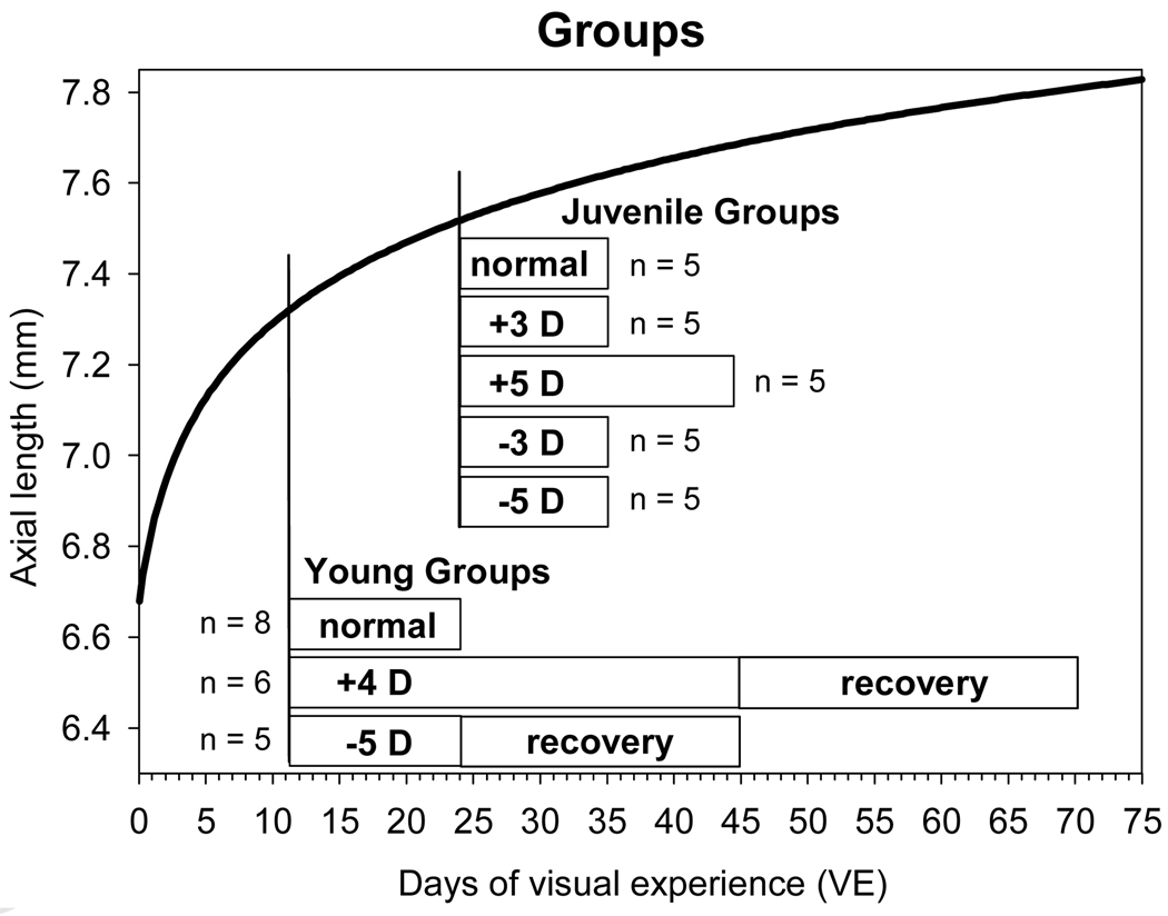
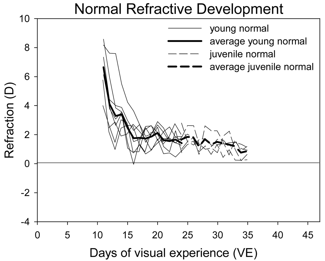
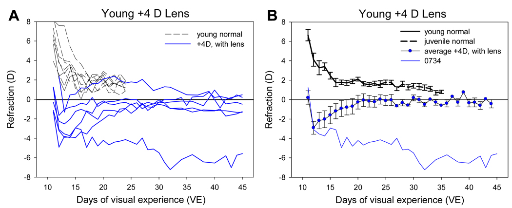
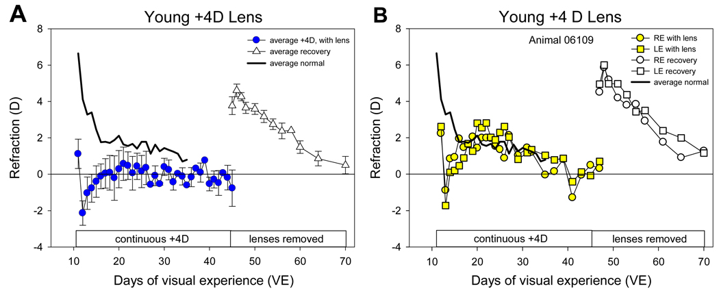
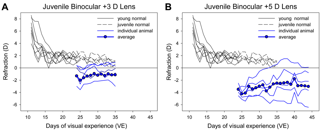
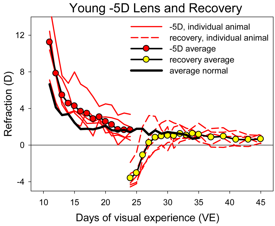
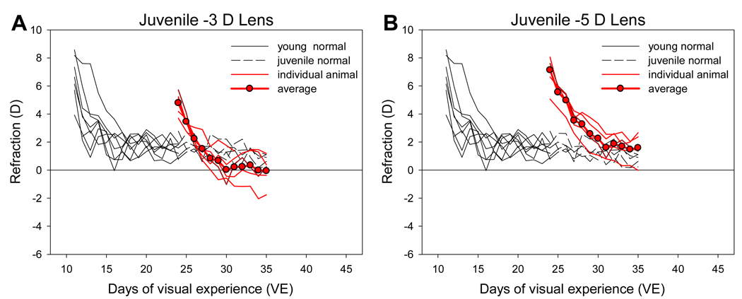
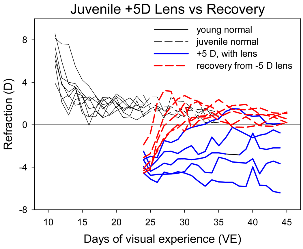
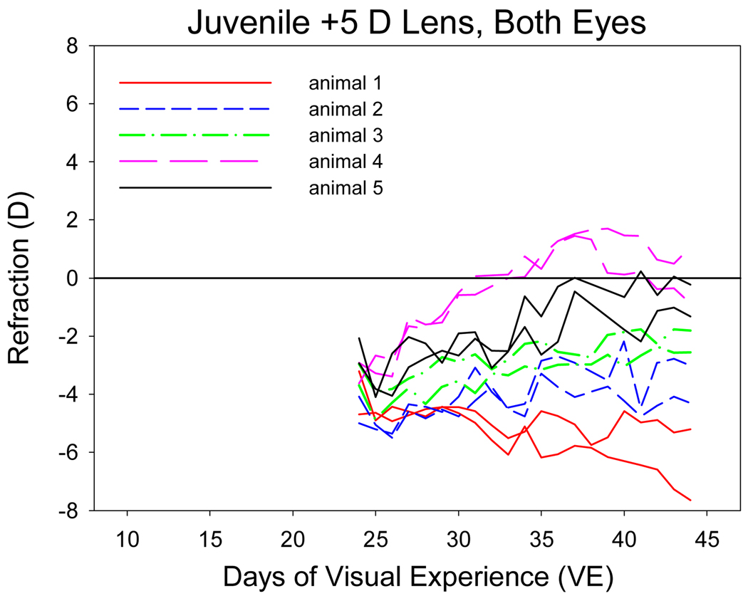
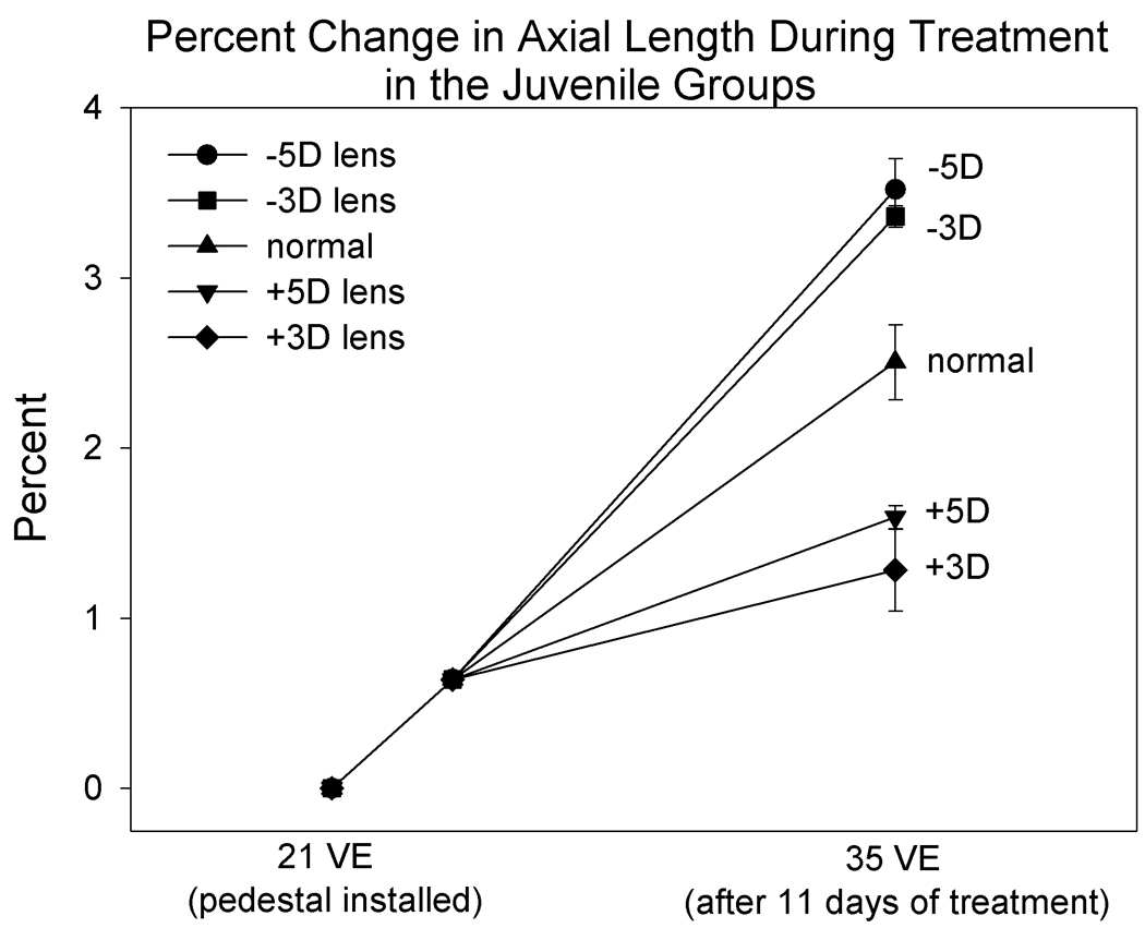
We examined normal emmetropization and the refractive responses to binocular plus or minus lenses in young (late infantile) and juvenile tree shrews. In addition, recovery from lens-induced myopia was compared with the response to a similar amount of myopia produced with plus lenses in age-matched juvenile animals. Normal emmetropization was examined with daily noncycloplegic autorefractor measures from 11 days after natural eye-opening (days of visual experience [VE]) when the eyes were in the infantile, rapid growth phase and their refractions were substantially hyperopic, to 35 days of VE when the eyes had entered the juvenile, slower growth phase and the refractions were near emmetropia. Starting at 11 days of VE, two groups of young tree shrews wore binocular +4 D lenses (n=6) or -5 D lenses (n=5). Starting at 24 days of VE, four groups of juvenile tree shrews (n=5 each) wore binocular +3 D, +5 D, -3 D, or -5 D lenses. Non-cycloplegic measures of refractive state were made frequently while the animals wore the assigned lenses. The refractive response of the juvenile plus-lens wearing animals was compared with the refractive recovery of an age-matched group of animals (n=5) that were myopic after wearing a -5 D lens from 11 to 24 days of VE. In normal tree shrews, refractions (corrected for the small eye artifact) declined rapidly from (mean±SEM) 6.6±0.6 D of hyperopia at 11 VE to 1.4±0.2 D at 24 VE and 0.8±0.4 D at 35 VE. Plus 4 D lens treatment applied at 11 days of VE initially corrected or over-corrected the young animals' hyperopia and produced a compensatory response in most animals; the eyes became nearly emmetropic while wearing the +4 D lenses. In contrast, plus-lens treatment starting at 24 days of VE initially made the juvenile eyes myopic (over-correction) and, on average, was less effective. The response ranged from no change in refractive state (eye continued to experience myopia) to full compensation (emmetropic with the lens in place). Minus-lens wear in both the young and juvenile groups, which initially made eyes more hyperopic, consistently produced compensation to the minus lens so that eyes reached age-appropriate refractions while wearing the lenses. When the minus lenses were removed, the eyes recovered quickly to age-matched normal values. The consistent recovery response from myopia in juvenile eyes after minus-lens compensation, compared with the highly variable response to plus lens wear in age-matched juvenile animals suggests that eyes retain the ability to detect the myopic refractive state, but there is an age-related decrease in the ability of normal eyes to use myopia to slow their elongation rate below normal. If juvenile human eyes, compared with infants, have a similar difficulty in using myopia to slow axial elongation, this may contribute to myopia development, especially in eyes with a genetic pre-disposition to elongate.
Copyright © 2010 Elsevier Ltd. All rights reserved.
Figures
References
-
- Andison ME, Sivak JG, Bird DM. The refractive development of the eye of the American kestrel (Falco sparverius): a new avian model. J Comp Physiol. 1992;170:565–574. - PubMed
-
- Bradley DV, Fernandes A, Lynn M, Tigges M, Boothe RG. Emmetropization in the rhesus monkey (Macaca mulatta): birth to young adulthood. Invest Ophthalmol. Vis. Sci. 1999;40:214–229. - PubMed
-
- Chung K, Mohidin N, O'Leary DJ. Undercorrection of myopia enhances rather than inhibits myopia progression. Vision Res. 2002;42:2555–2559. - PubMed
-
- Cook RC, Glasscock RE. Refractive and ocular findings in the newborn. Am J Ophthalmol. 1951;34:1407–1413. - PubMed
-
- Cottriall CL, McBrien NA. The M1 muscarinic antagonist pirenzepine reduces myopia and eye enlargement in the tree shrew. Invest. Ophthalmol. Vis. Sci. 1996;37:1368–1379. - PubMed
Publication types
MeSH terms
Grants and funding
LinkOut - more resources
Full Text Sources
Other Literature Sources
Medical
