. 2010 Nov;42(11):1864-75.
doi: 10.1016/j.biocel.2010.08.004. Epub 2010 Aug 14.

Adjudin-mediated Sertoli-germ cell junction disassembly affects Sertoli cell barrier function in vitro and in vivo

Affiliations

Adjudin-mediated Sertoli-germ cell junction disassembly affects Sertoli cell barrier function in vitro and in vivo

Linlin Su et al. Int J Biochem Cell Biol. 2010 Nov.

Abstract

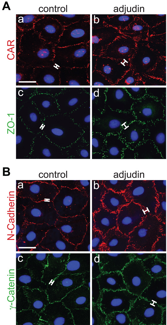
Adjudin, an analogue of lonidamine, affects adhesion between Sertoli and most germ cells, resulting in reversible infertility in rats, rabbits and dogs. Previous studies have described the apical ectoplasmic specialization, a hybrid-type of Sertoli cell-elongating/elongated spermatid adhesive junction, as a key target of adjudin. In this study, we ask if the function of the blood-testis barrier which is constituted by co-existing tight junctions, desmosome-gap junctions and basal ectoplasmic specializations can be maintained when the seminiferous epithelium is under assault by adjudin. We report herein that administration of a single oral dose of adjudin to adult rats increased the levels of several tight junction and basal ectoplasmic specialization proteins during germ cell loss from the seminiferous epithelium. These findings were corroborated by a functional in vitro experiment when Sertoli cells were cultured on Matrigel™-coated bicameral units in the presence of adjudin and transepithelial electrical resistance was quantified across the epithelium. Indeed, the Sertoli cell permeability barrier was shown to become tighter after adjudin treatment as evidenced by an increase in transepithelial electrical resistance. Equally important, the blood-testis barrier in adjudin-treated rats was shown to be intact 2 weeks post-treatment when its integrity was monitored following vascular administration of inulin-fluorescein isothiocyanate which failed to permeate past the barrier and enter into the adluminal compartment. These results illustrate that a unique mechanism exists to maintain blood-testis barrier integrity at all costs, irrespective of the presence of germ cells in the seminiferous epithelium of the testis.


Figures

Fig. 1
Fig. 1. Adjudin-mediated Sertoli–germ cell junction disassembly up-regulates BTB-constituent proteins in vivo
Adjudin (50 mg/kg b.w.) was administered to rats (~300 g b.w., n = 4/time point) as described in Materials and methods. Control rats (n = 3) received 0.5% methyl-cellulose [wt/vol] only. (A) Immunoblots investigating changes in TJ and basal ES proteins in the testis following oral administration of adjudin. Actin served as a loading control. (Ba, b) Histograms summarizing results shown in (A). Each data point was normalized against its corresponding actin data point and then compared against its control [0 day (D)] which was arbitrarily set at 1. Error bars represent mean ± SD of data using testis lysates from 4 different animals. *, P<0.05; **, P<0.01 (Student’s t-test).
Fig. 2
Fig. 2. Adjudin affects protein localization at the BTB
Adjudin (50 mg/kg b.w.) was administered to rats (~300 g b.w., n = 4/time point) as described in Materials and methods. Control rats (n = 3) received 0.5% methyl-cellulose [wt/vol] only. Cross-sections of testes from control and adjudin-treated rats at 7 and 14 days post-treatment immunostained for claudin-11 (A–C), occludin (D–F), JAM-A (G–I), CAR (J–L), N-cadherin (M–O) and γ-catenin (P–R). Arrowheads (J) indicate the presence of CAR at the apical ES. Brackets show a thickening in claudin-11 (B, C versus A), occludin (E, F versus D), JAM-A (H, I versus G), CAR (K, L versus J), N-cadherin (N, O versus M) and γ-catenin (Q, R, versus P) immunostaining at the BTB following adjudin treatment when compared to control rats. This experiment was repeated three times using testes from three different rats treated with adjudin. Bar (A, also corresponds to B–R) = 115 µm.
Fig. 3
Fig. 3. Adjudin strengthens Sertoli cell barrier function in vitro and displays no cytotoxicity
(A–C) TER was measured as a means to assess barrier function when Sertoli cells were cultured in the presence of adjudin. (A) Sertoli cells were cultured at high density on Matrigel™-coated bicameral units. Thereafter, increasing concentrations of adjudin (100 ng, 500 ng and 1 µg/ml) were added to both the apical and basal compartment of bicameral units onwards of day 4. (B, C) Adjudin (500 ng/ml) was added to the apical (B) and/or the basal (C, see schematic drawing in B and C) compartment of bicameral units onwards of day 4. Media containing the appropriate concentration of adjudin was replaced daily, and TER readings were recorded every 12 hr. Results in (A–C) are representative of at least four independent experiments using different batches of isolated Sertoli cells, and one set of results is shown in (A–C). Each adjudin treatment data point in (A–C) was compared to its corresponding control (i.e., Sertoli cells cultured in the presence of vehicle which was either added to both compartments, the apical or basal compartment alone). The addition of adjudin to both the apical and basal compartment of bicameral units (B, C) served as an internal control. Results in (B, C) represent data from a single experiment, but these results were presented as two separate plots. Error bars represent mean ± SD of triplicate bicameral units per treatment group in a single experiment. *, P<0.05; **, P<0.01 (ANOVA followed by Dunnett’s test). (D) Cytotoxicity assay to assess if adjudin displays overt toxicity in Sertoli cells. Sertoli cells, cultured at high density on Matrigel™-coated plates for 4 days, were treated with increasing concentrations of adjudin (0.001 to 500 µg/ml) for 3 days, followed by addition of using XTT reagent for 6 hours. The dotted-line indicates the maximum dose of adjudin that was used for all in vitro experiments. This cytotoxicity assay was repeated three times using different batches of isolated Sertoli cells treated with increasing doses of adjudin.
Fig. 4
Fig. 4. Adjudin up-regulates Sertoli cell barrier constituent proteins in vitro
Sertoli cells were cultured at high density on Matrigel™-coated dishes as described in Materials and methods. On day 4, adjudin was added (this time point was designated as 0 hour), and cells were used at different time points thereafter for lysate preparation. Control Sertoli cells consisted of adding vehicle alone. (A) Immunoblots investigating changes in TJ and basal ES proteins following treatment of Sertoli cells with adjudin (500 ng and/or 1 µg/ml). Actin served as a loading control. (Ba–f) Histograms summarizing results shown in (A). Each data point was normalized against its corresponding actin data point. Error bars represent mean ± SD of data from at least three different batches of isolated Sertoli cells treated with adjudin. *, P<0.05; **, P<0.01 (ANOVA followed by Dunnett’s test).
Fig. 5
Fig. 5. Adjudin affects protein localization at the Sertoli cell surface
Sertoli cells were cultured at high density on Matrigel™-coated glass coverslips as described in Materials and methods. On day 4, adjudin was added. Three days thereafter, cells were used for immunofluorescent microscopy. (A, B) Control and adjudin-treated Sertoli cells were immunostained for CAR (Aa, b), ZO-1 (Ac, d), N-cadherin (Ba, b) and γ-catenin (Bc, d). Brackets show a thickening in CAR (Ab versus a), ZO-1 (Ad versus c), N-cadherin (Bb versus a) and γ-catenin (Bd versus c) immunostaining at the Sertoli cell interface following adjudin treatment. This immunofluorescent microscopy experiment was repeated at least three times using different batches of isolated Sertoli cells treated with adjudin. Bar (Aa, also corresponds to b–d; Ba, also corresponds to b–d) = 15 µm.
Fig. 6
Fig. 6. Changes in F-actin at the BTB after administration of adjudin in vivo
Adjudin (50 mg/kg b.w.) was administered to rats (~300 g b.w., n = 4/time point) as described in Materials and methods, and the integrity of F-actin at the BTB was assessed by using rhodamine phalloidin and fluorescent optics. Control rats (n = 3) received 0.5% methyl-cellulose [wt/vol] only. (A–D) Representative testis cross-section from a control rat. (E–L) Representative testis cross-sections from rats 7 (E–H) and 14 (I–L) days after administration of adjudin. Dotted-line boxes in (A, B, E, F, I, K) represent magnified views. Arrowheads (C, G, K) indicate changes in F-actin orientation at the BTB. Brackets (K versus C) indicate a thickening in F-actin after administration of adjudin when compared to the control. Nuclei were visualized by using mounting medium containing DAPI (D, H, L) which served as an indicator of germ cell loss from seminiferous tubules. Dotted-line circles (D, H, L) outline the periphery of seminiferous tubules. bv, blood vessel. Bar (A, also corresponds to E, I) = 175 µm; bar (B, also corresponds to D, F, H, J, L) = 110 µm; bar (C, also corresponds to G, K) = 80 µm.
Fig. 7
Fig. 7. Adjudin-mediated Sertoli-germ cell junction disassembly does not compromise barrier function in vivo
Inulin-FITC was administered to control and treated rats (~300 g b.w., n = 3/time point) as described in Materials and methods, and testis cryo-sections were examined microscopically by fluorescent optics. (A) Control rats (n = 3) in the adjudin group received 0.5% methyl-cellulose [wt/vol], whereas control rats (n = 3) in the CdCl2 group received PBS (data not shown). (B) Representative testis cross-section from a rat 5 days (D) after administration of CdCl2 (3 mg/kg b.w.). (C–E) Representative testis cross-sections from rats 4 (C), 7 (D) and 14 (E) days after administration of adjudin (50 mg/kg b.w.). Dotted-line circles (A–E) outline the periphery of seminiferous tubules. Brackets (A–E) show the relative distance that inulin-FITC had traveled from the periphery of seminiferous tubules. bv, blood vessel. Bar (A, also corresponds to B–E) = 115 µm.

References

    1. Baillie AH, Griffiths K. 3β-Hydroxysteroid dehydrogenase activity in the mouse Leydig cell. J Endocrinol. 1964;29:9–17. - PubMed
    1. Balda MS, Matter K. Tight junctions and the regulation of gene expression. Biochim Biophys Acta. 2009;1788:761–767. - PubMed
    1. Beaver BV, Reed W, Leary S, McKiernan B, Bain F, Schultz R, et al. 2000 Report of the American Veterinary Medical Association Panel in Euthanasia. J Am Vet Med Assoc. 2001;218:669–696. - PubMed
    1. Ben-Ze'ev A, Geiger B. Differential molecular interactions of β-catenin and plakoglobin in adhesion, signaling, and cancer. Curr Opin Cell Biol. 1998;10:629–639. - PubMed
    1. Byers S, Hadley MA, Djakiew D, Dym M. Growth and characterization of epididymal epithelial cells and Sertoli cells in dual environment culture chambers. J Androl. 1986;7:59–68. - PubMed

Publication types

MeSH terms

LinkOut - more resources