

Endolymphatic hydrops: pathophysiology and experimental models
- PMID: 20713237
- PMCID: PMC2923478
- DOI: 10.1016/j.otc.2010.05.007
Endolymphatic hydrops: pathophysiology and experimental models
Abstract
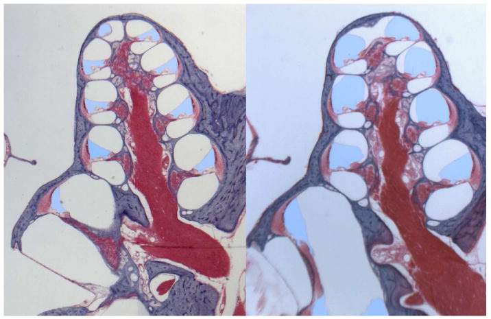
It is well established that endolymphatic hydrops plays a role in Ménière disease, even though the precise role is not fully understood and the presence of hydrops in the ear does not always result in symptoms of the disease. It nevertheless follows that a scientific understanding of how hydrops arises, how it affects the function of the ear, and how it can be manipulated or reversed could contribute to the development of effective treatments for the disease. Measurements in animal models in which endolymphatic hydrops has been induced have given numerous insights into the relationships between hydrops and other pathologic and electrophysiological changes, and how these changes influence the function of the ear. The prominent role of the endolymphatic sac in endolymph volume regulation, and the cascade of histopathological and electrophysiological changes that are associated with chronic endolymphatic hydrops, have now been established. An increasing number of models are now available that allow specific aspects of the interrelationships to be studied. The yclical nature of Ménière symptoms gives hope that treatments can be developed to maintain the ear in permanent state of remission, possibly by controlling endolymphatic hydrops, thereby avoiding the rogressive damage and secondary pathologic changes that may also contribute to the patient's symptoms.
Copyright 2010 Elsevier Ltd. All rights reserved.
Figures
References
-
- Yamakawa K. Über die pathologische Veränderung beieinem Meniere-Kranken. J Otolaryngol Soc Japan; Proc 42nd Ann Meet Oto-Rhino-Laryngol Soc Japan; 1938. pp. 2310–2312.
-
- Kimura RS, Schuknecht HF. Membranous hydrops in the inner ear of the guinea pig after obliteration of the endolymphatic sac. Pract Oto-rhino-laryng. 1965;27:343–354.
-
- Kimura RS. Experimental blockage of the endolymphatic duct and sac and its effect on the inner ear of the guinea pig. Ann Oto-Rhino-laryng. 1967;76:664–687. - PubMed
-
- Salt AN, DeMott JE, Kimura RS. Comparison of endolymph cross-sectional area measured histologically with that measured in vivo with an ionic volume marker. Ann Otol Rhinol Laryngol. 1995b;104:886–894. - PubMed
Publication types
MeSH terms
Substances
Grants and funding
LinkOut - more resources
Full Text Sources
Other Literature Sources
