

Genetic and transcriptional control of bone formation
- PMID: 20713262
- PMCID: PMC2923651
- DOI: 10.1016/j.coms.2010.05.001
Genetic and transcriptional control of bone formation
Abstract
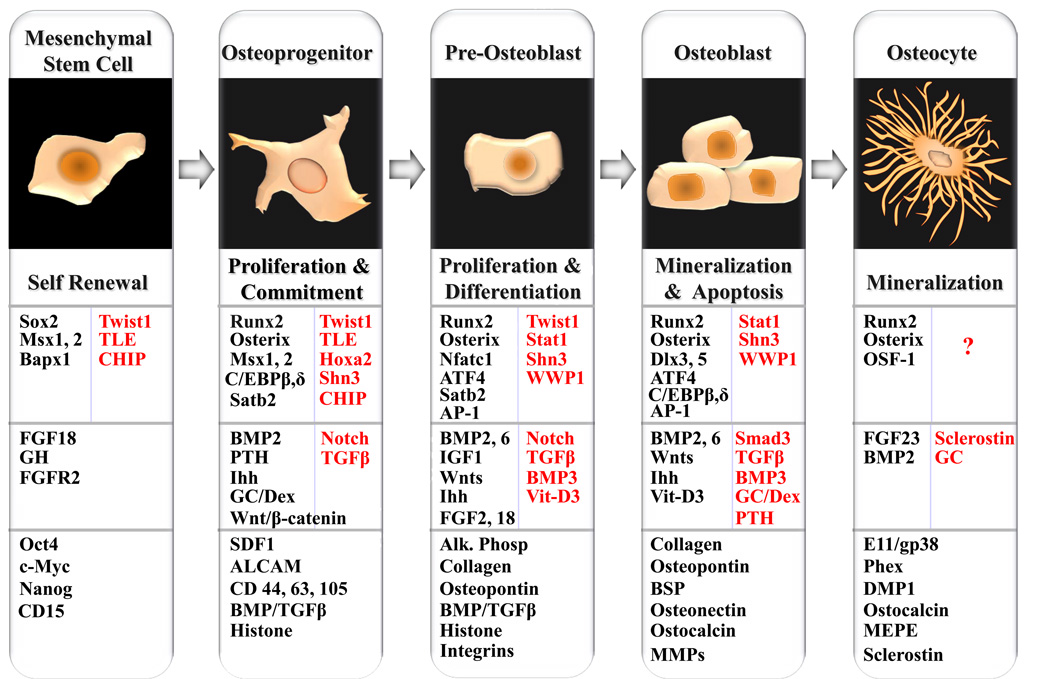
An exquisite interplay of developmental cues, transcription factors, and coregulatory and signaling proteins support formation of skeletal elements of the jaw during embryogenesis and dynamic remodeling of alveolar bone in postnatal life. These molecules promote initial condensation of the mesenchyme, commitment of the mesenchymal progenitor to osteogenic lineage cells, and differentiation of committed osteoblasts to mature osteocytes within mineralized bone. Parallel regulatory networks promote formation of the functional osteoclast from mononuclear cells to support continuous bone remodeling within the alveolar bone. With an ever expanding list of new regulatory factors, the complexities of the molecular mechanisms that control gene expression in skeletal cells are being further appreciated. This article examines the multifunctional roles of prominent nuclear proteins, cytokines, hormones, and paracrine factors that control osteogenesis.
Copyright 2010 Elsevier Inc. All rights reserved.
Figures
References
-
- Shubin NH, Alberch PA. Morphogenic approach to the origin and basic organization of the tetrapod limb. Evol. Biol. 1986;20:319–387.
-
- Osumi-Yamashita N, Ninomiya Y, Eto K, et al. The contribution of both forebrain and midbrain crest cells to the mesenchyme in the frontonasal mass of mouse embryos. Dev. Biol. 1994;164(2):409–419. - PubMed
-
- Sodek J, McKee MD. Molecular and cellular biology of alveolar bone. Periodontology. 2000;2000(24):99–126. - PubMed
-
- Zernik JH, Nowroozi N, Liu YH, et al. Development, maturation, and aging of the alveolar bone. New insights. Dent Clin North Am. 1997;41(1):1–15. - PubMed
-
- Chai Y, Jiang X, Ito Y, et al. Fate of the mammalian cranial neural crest during tooth and mandibular morphogenesis. Development. 2000;127:1671–1679. - PubMed
Publication types
MeSH terms
Substances
Grants and funding
LinkOut - more resources
Full Text Sources
Other Literature Sources
