

Review
doi: 10.1102/1470-7330.2010.0026.
Primary and secondary neoplasms of the spleen
Affiliations
- PMID: 20713317
- PMCID: PMC2943678
- DOI: 10.1102/1470-7330.2010.0026
Item in Clipboard
Review
Primary and secondary neoplasms of the spleen
Cancer Imaging.
.
Abstract
With the exception of lymphoma involving the spleen, other primary and secondary neoplasms are rare and infrequently encountered. Primary malignant neoplasms involving the spleen are lymphoma and angiosarcoma. Primary benign neoplasms involving the spleen include hemangioma, lymphangioma, littoral cell angioma and splenic cyst and solid lesions such as hamartoma and inflammatory pseudotumor.
Figures
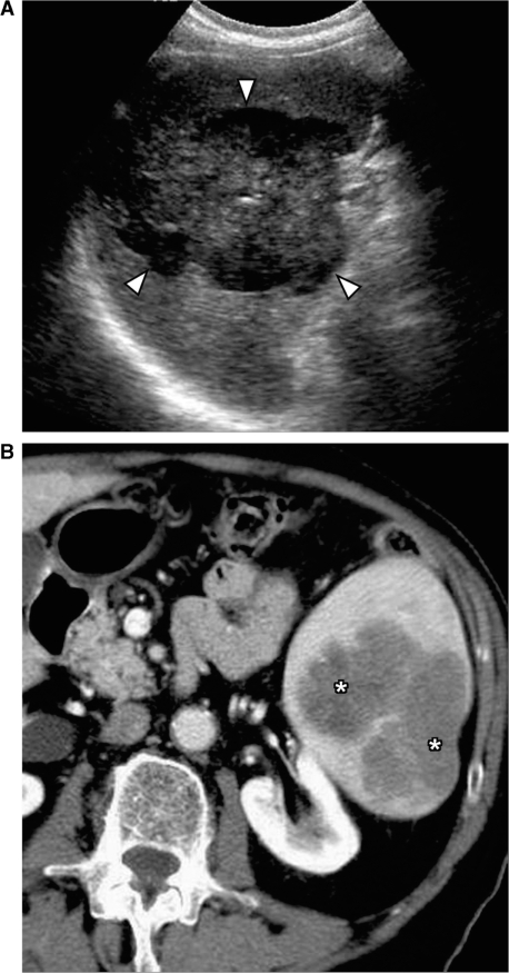
Primary splenic lymphoma. (A) Ultrasound shows a large heterogeneous mass in the spleen (arrowheads). (B) Contrast-enhanced CT shows a lobulated hypodense mass in the spleen (*). Biopsy confirmed it to be non-Hodgkin lymphoma of B-cell origin.
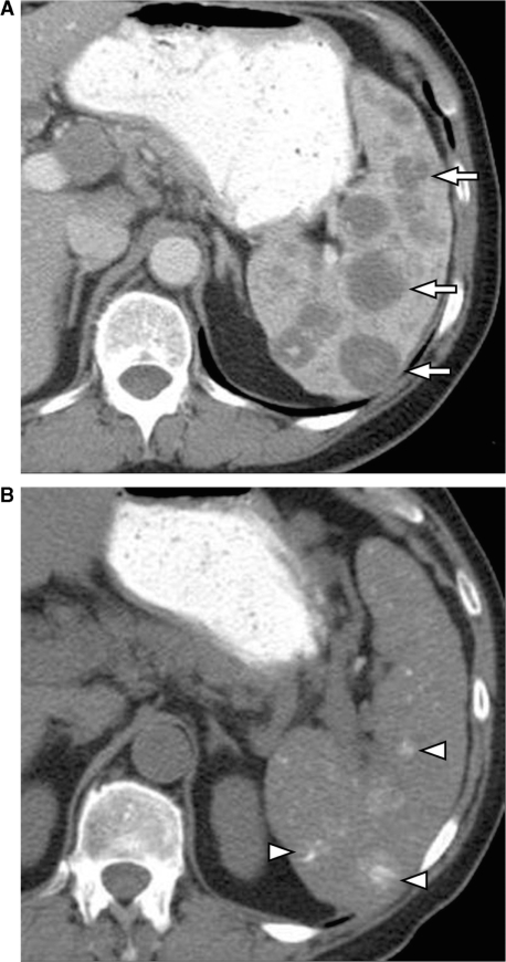
Calcification in the spleen following treatment for lymphoma. (A) Contrast-enhanced CT shows multiple hypodense lesions in the spleen (arrows) secondary to lymphomatous deposits. (B) Unenhanced CT scan obtained 2 years after chemotherapy shows multiple foci of calcification representing treated lymphoma (arrowheads).
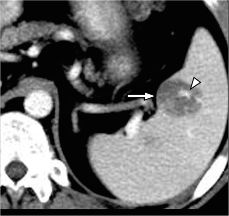
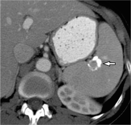
Splenic angiosarcoma. Contrast-enhanced CT shows a hypodense mass in the spleen (arrow) with focal areas of enhancement secondary to the vascular nature of tumor (arrowhead).
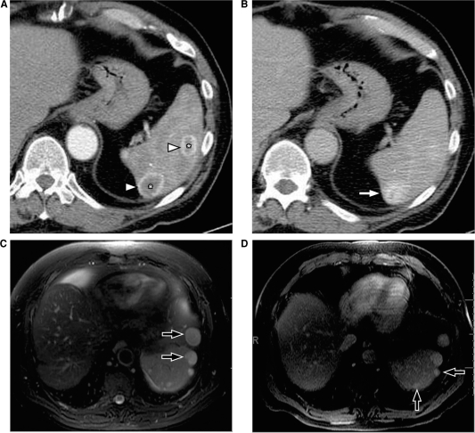
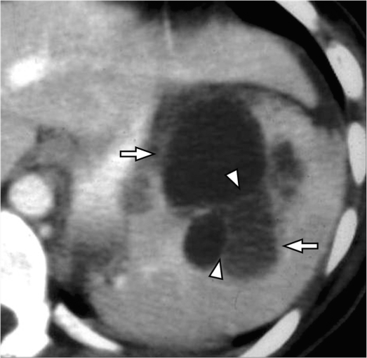
Splenic hemangiomas. (A) Arterial phase of contrast-enhanced CT shows two lesions in the spleen (*), which demonstrate the peripheral continuous rim of enhancement (arrowheads). (B) Venous phase shows progressive centripetal enhancement in both of the lesions, which is better seen in the posterior lesion (arrow). MRI in a different patient shows multiple hemangiomas (arrows) which are hyperintense on T2-weighted images (C) and show retention of contrast on delayed post contrast T1-weighted images (D).
Splenic lymphangioma. Contrast-enhanced CT shows a large multicystic lesion (arrows) with thin septations (arrowheads) in the spleen extending to the capsular margin.
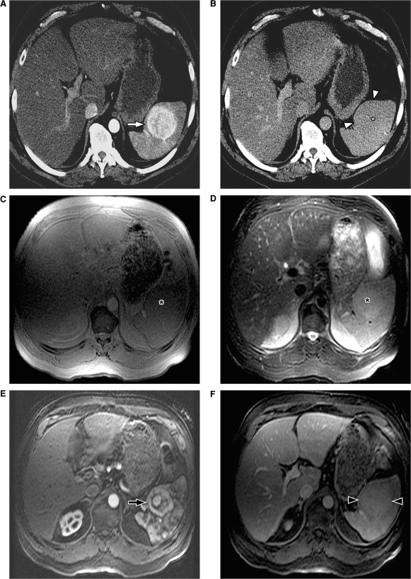
Splenic hamartoma. (A) Arterial phase of contrast-enhanced CT shows an enhancing mass in the spleen (arrow). (B) The mass is isodense on the venous phase (asterisk) and can be appreciated only as a capsular bulge (arrowheads). On MRI, the mass is isointense on the T1-weighted inphase (C) and is slightly hyperintense on the T2-weighted fat-saturated sequence (asterisk) (D). After gadolinium administration, heterogeneous enhancement is noted in the arterial phase (arrow) (E) with uniform delayed enhancement noted on the delayed phase (arrowheads) (F).

Littoral cell angioma of the spleen. (A,B) Contrast-enhanced axial CT images of the spleen show multiple well-defined hypodense lesions (arrowheads) ranging in size from 5 mm to 3 cm. Pathology showed littoral cell angioma of the spleen.
Splenic sarcoma. Contrast-enhanced CT shows a large exophytic mass arising from the spleen (arrows). Large necrotic areas (*) and few foci of calcification (arrowhead) are noted within the lesion.
Splenic metastasis from melanoma. Contrast-enhanced CT shows multiple hypodense masses in the liver (arrowheads) and a large mass in the spleen (arrow) demonstrating central necrosis (*) in a patient with known melanoma representing hepatic and splenic metastasis from melanoma.
Calcified splenic metastasis from ovarian cancer. Contrast-enhanced CT shows a heterogeneously calcified lesion in the spleen (arrow) in a patient with treated ovarian cancer metastasis.

Perisplenic deposits in pseudomyxoma peritonei. Contrast-enhanced CT scan shows multiple well-defined cystic lesions in the perisplenic region invaginating into the spleen (arrows) in a patient with known pseudomyxoma peritonei. Similar cystic deposits are also noted in the perigastric region (*).
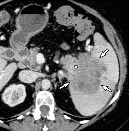
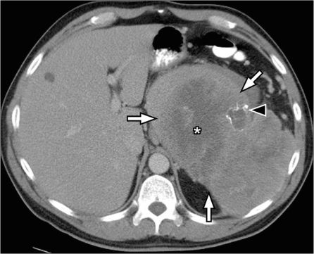
Pancreatic cancer invading through the splenic hilum. Contrast-enhanced CT shows a large heterogeneous mass centered on the splenic hilum (arrows) and inseparable from the tail of the pancreas (*) representing pancreatic adenocarcinoma of the pancreatic tail with splenic invasion.

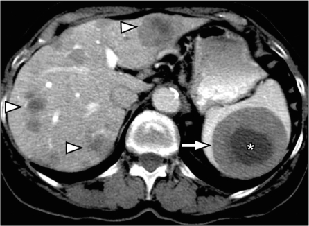
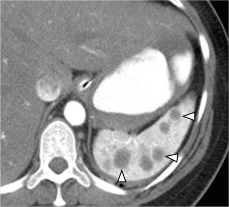
Splenic sarcoidosis. Contrast-enhanced CT shows splenomegaly with innumerable hypodense nodules in the spleen (white arrowheads). Multiple tiny nodules in the liver (black arrowheads) and few enlarged lymph nodes along the celiac axis (arrow) are noted in a patient with known sarcoidosis representing hepatosplenic involvement and lymph node enlargement.
Peliosis of spleen. Contrast-enhanced CT shows multiple hypodense lesions (arrowheads) in a normal-sized spleen. Pathology showed peliosis of the spleen.
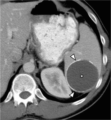
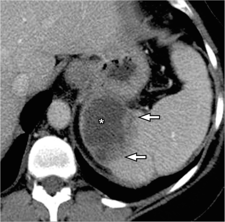
Splenic pseudocyst. Contrast-enhanced CT shows a well-defined cyst in the spleen (*) with peripheral rim of calcification (arrowheads) and no internal septations, presumed to be a post-traumatic pseudocyst.
Splenic abscess. Contrast-enhanced CT shows a heterogeneous cystic lesion (*) with thick irregular enhancing margins (arrows) in a patient with septicemia representing a splenic abscess.
References
-
- Dachman A. Radiology of the spleen. St. Louis, MO: Mosby; 1993.
-
- Kamaya A, Weinstein S, Desser TS. Multiple lesions of the spleen: differential diagnosis of cystic and solid lesions. Semin Ultrasound CT MR. 2006;27:389–403. doi:10.1053/j.sult.2006.06.004. - DOI - PubMed
-
- Spier CM, Kjeldsberg CR, Eyre HJ, Behm FG. Malignant lymphoma with primary presentation in the spleen. A study of 20 patients. Arch Pathol Lab Med. 1985;109:1076–80. - PubMed
-
- Bhatia K, Sahdev A, Reznek RH. Lymphoma of the spleen. Semin Ultrasound CT MR. 2007;28:12–20. doi:10.1053/j.sult.2006.10.010. - DOI - PubMed
-
- Ahmann DL, Kiely JM, Harrison EG, Jr, Payne WS. Malignant lymphoma of the spleen. A review of 49 cases in which the diagnosis was made at splenectomy. Cancer. 1966;19:461–9. doi:10.1002/1097-0142(196604)19:4<461::AID-CNCR2820190402>3.0.CO;2-X. - DOI - PubMed
Publication types
MeSH terms
LinkOut - more resources
Full Text Sources
Medical