. 2010 Oct 22;285(43):33037-33044.
doi: 10.1074/jbc.M110.155283. Epub 2010 Aug 16.

Characterization of hMTr1, a human Cap1 2'-O-ribose methyltransferase

Affiliations

Characterization of hMTr1, a human Cap1 2'-O-ribose methyltransferase

François Bélanger et al. J Biol Chem. .

Abstract

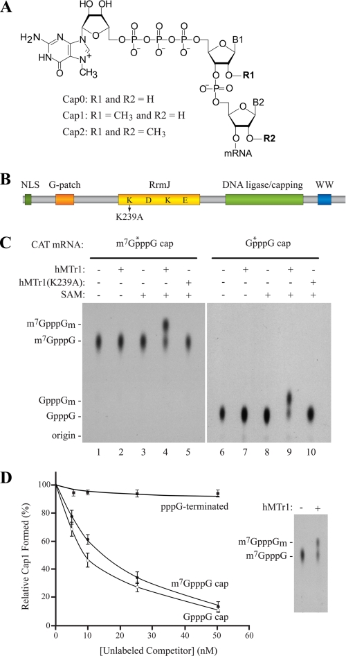
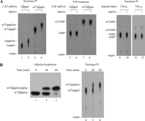
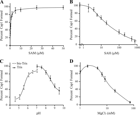
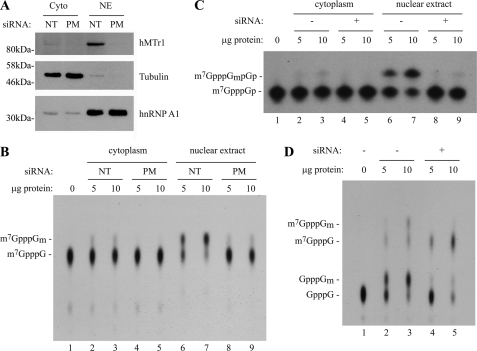
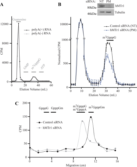
Cellular eukaryotic mRNAs are capped at their 5' ends with a 7-methylguanosine nucleotide, a structural feature that has been shown to be important for conferring mRNA stability, stimulating mRNA biogenesis (splicing, poly(A) addition, nucleocytoplasmic transport), and increasing translational efficiency. Whereas yeast mRNAs have no additional modifications to the cap, called cap0, higher eukaryotes are methylated at the 2'-O-ribose of the first or the first and second transcribed nucleotides, called cap1 and cap2, respectively. In the present study, we identify the methyltransferase responsible for cap1 formation in human cells, which we call hMTr1 (also known as FTSJD2 and ISG95). We show in vitro that hMTr1 catalyzes specific methylation of the 2'-O-ribose of the first nucleotide of a capped RNA transcript. Using siRNA-mediated knockdown of hMTr1 in HeLa cells, we demonstrate that hMTr1 is responsible for cap1 formation in vivo.


Figures

FIGURE 1.
FIGURE 1.
hMTr1 methylates capped mRNA in vitro. A, structure of the 5′-cap in eukaryotic mRNA. B, schematic representation of hMTr1. Predicted domains include a nuclear localization signal (NLS), a G patch RNA interaction domain, am RrmJ homology 2′-O-ribose methyltransferase domain, a putative DNA ligase/capping domain, and a WW protein interaction domain. K239 denotes a point mutation in the conserved KDKE catalytic tetrad. C, hMTr1-methylated mRNAs containing m7GpppG or GpppG cap structures. In vitro transcribed CAT mRNA was capped with [α-32P]GTP and treated with purified recombinant hMTr1 (wild type or K239A mutant) in the presence or absence of 50 μm SAM. Nuclease P1-digested products were analyzed by TLC. m7GpppG- (lanes 1–5) and GpppG- (lanes 6–10) terminated CAT mRNA were used as substrate. *, positions of the radiolabeled phosphate. Unlabeled cap structure analogs were analyzed in parallel, and their positions of migration were detected by UV shadowing and are indicated on the left. D, methylation of RNA by hMTr1 requiring a cap structure. Reactions were performed as in C, with the indicated amounts of unlabeled CAT mRNA competitors. The inset shows that the efficiency of conversion of m7GpppG to m7GpppGm was ∼50% in the absence of competitor.
FIGURE 2.
FIGURE 2.
hMTr1 methylates the 2′-O-ribose of the first transcribed nucleotide. A, hMTr1 does not methylate the terminal m7G moiety. Products from in vitro methylation reactions were treated either with nuclease P1 (lanes 1–4) or with tobacco acid pyrophosphatase (TAP; lanes 5–8) to release the terminal [32P]m7GMP or [32P]GMP. Internally labeled CAT mRNA was also used as substrate (lanes 9–12). Products were analyzed by TLC. Migration of unlabeled markers are indicated on the left. B, methylation of the first transcribed nucleotide by hMTr1 occurs on the 2′-O-ribose. Methylation reactions were performed for the indicated time, and products were degraded by complete alkaline hydrolysis and analyzed on a 25% PAGE-urea (lanes 1–3). Digestion of the products by nuclease P1 followed by TLC was performed in parallel (lanes 4–6).
FIGURE 3.
FIGURE 3.
Biochemical characterization of hMTr1. A, activity of hMTr1 is dependent on SAM concentration. Reactions were performed with increasing SAM concentrations. Signals corresponding to cap0 and cap1 were quantified and expressed as a percentage of cap1 formed. B, activity of hMTr1 is inhibited by S-adenosylhomocysteine (SAH). Methylation reactions were performed in the presence of 5 μm SAM and increasing concentrations of S-adenosylhomocysteine. C, pH affects hMTr1 activity. Reactions were performed at the indicated pH in either Tris (filled circles) or bis-Tris (open circles). D, magnesium affects hMTr1 methyltransferase activity.
FIGURE 4.
FIGURE 4.
Knockdown of hMTr1 in HeLa cells results in loss of cap1 methyltransferase activity. A, Western blots document subcellular fractionation and efficiency of hMTr1 knockdown. HeLa cells were treated with perfect match siRNAs directed against hMTr1 (PM) or with nontargeting control siRNAs (NT) and fractionated into cytoplasmic (Cyto) and nuclear extracts (NE). Western blotting was to detect the presence of hMTr1, tubulin (cytoplasmic), and hnRNP A1 (predominantly nuclear). B, extracts prepared from hMTr1 knockdown cells have reduced 2′-O-methyltransferase activity. Radiolabeled m7GpppG-terminated CAT mRNA was incubated in the presence of HeLa cytoplasmic or nuclear extracts. Formation of m7GpppGm was assessed by nuclease P1 digestion of the reaction products followed by TLC analysis. C, extracts prepared from hMTr1 knockdown cells have reduced 2′-O-methyltransferase activity. Reactions were the same as in B, except the reaction products were analyzed by alkaline hydrolysis and electrophoretic separation of the products on a 25% polyacrylamide-8 m urea gel. D, loss of 2′-O-methylation does not affect conversion of GpppG to m7GpppG. Radiolabeled GpppG-terminated CAT mRNA was incubated in the presence of HeLa nuclear extracts, and reaction products were digested with nuclease P1 and analyzed by TLC.
FIGURE 5.
FIGURE 5.
hMTr1 is responsible for Cap1 formation on endogenous mRNAs in vivo. A, HeLa cells were labeled with l-[methyl-3H]methionine. Purified poly(A)+ mRNA and poly(A) RNAs were treated with nuclease P1 followed by antartic phosphatase and analyzed by DEAE anion exchange chromatography. Gray bars represent the fractions in which unlabeled markers eluted (guanosine, GMP, m7GpppG, and ATP). B, knockdown of hMTr1 decreases incorporation of methyl-3H in the cap structure. HeLa cells were transfected with hMTr1 perfect match siRNA (PM) or a nontargeting control siRNA (NT) before labeling and DEAE analysis. Counts of each samples are normalized to an equivalent amount of total radioactivity. Values are average from three independent experiments. The inset is a Western blot showing the knockdown efficiency of hMTr1. C, knockdown of hMTr1 results in loss of cap1 2′-O-methylation of endogenous mRNAs. Fractions co-eluting with m7GpppG from B were combined and analyzed by TLC. Lanes were cut into 1.5-cm pieces and quantitated by scintillation counting. Horizontal bars denote the positions of various markers.

Similar articles

Cited by

References

    1. Shatkin A. J. (1976) Cell 9, 645–653 - PubMed
    1. Gu M., Lima C. D. (2005) Curr. Opin. Struct. Biol. 15, 99–106 - PubMed
    1. Shuman S. (2002) Nat. Rev. Mol. Cell. Biol. 3, 619–625 - PubMed
    1. Ensinger M. J., Moss B. (1976) J. Biol. Chem. 251, 5283–5291 - PubMed
    1. Venkatesan S., Gershowitz A., Moss B. (1980) J. Biol. Chem. 255, 2829–2834 - PubMed

Publication types